
- Title
-
Dorsal and intermediate neuronal cell types of the spinal cord are established by a BMP signaling pathway
- Authors
- Nguyen, V.H., Trout, J., Connors, SA.., Andermann, P., Weinberg, E., and Mullins, M.C.
- Source
- Full text @ Development
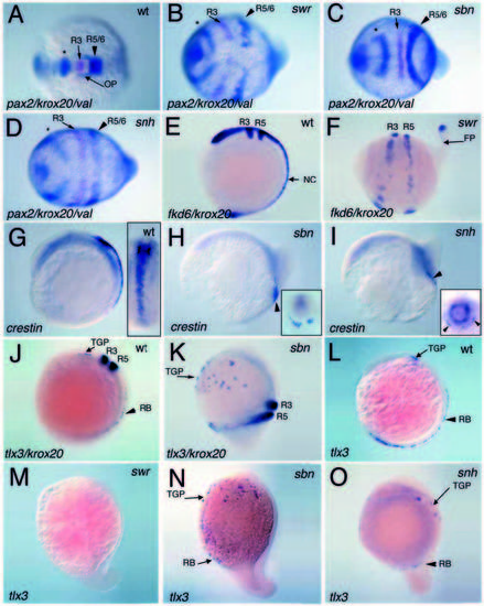
Conserved anteroposterior neural patterning (A-D) and loss of trunk neural crest (E-I) and dorsal RB neurons (J-O) in dorsalized mutant embryos. (A-D) are two-color triple in situ hybridizations in 6- somite-stage wild-type (A), swr/bmp2b (B), sbn/smad5 (C) and snh/bmp7 (D) embryos (dorsal views with anterior to the left). Expression of pax2.1 in the midbrain-hindbrain boundary (marked with an *) and val in rhombomeres 5 and 6 are in blue, while krox20 expression in rhombomere 3 is in magenta. Similar results were found in 1- to 2-somitestage embryos. Expression of fkd6 and krox20 in wild-type (E) and a swr/bmp2b mutant (F) at the 6- somite stage. crestin expression posterior to rhombomere 5 and within the rostral trunk of a 9-somitestage wild-type embryo (G), and in a greatly reduced domain in sbn/smad5 (H) and snh/bmp7 (I) mutant embryos. Insets in G-I are dorsal or posterior views. Expression of tlx3 and krox20 in 7-somite-stage wildtype (J) and sbn/smad5 (K) embryos. tlx3 expression in 7-somite wild-type (L), swr/bmp2b (M), sbn/smad5 (N) and snh/bmp7 (O) mutant embryos. In 0/8 swr/bmp2b, 8/20 sbn/smad5 and 5/8 snh/bmp7 mutant embryos, some RB-tlx3 expressing cells were detected, but in severely reduced numbers. Lateral views (E-O) with anterior to the top, dorsal to the right in E,G,J-O, and anterior to the left, dorsal to the top in F,H,I. R3, rhombomere 3; R5, rhombomere 5; R5/6, rhombomeres 5 and 6; OP, otic placode; NC, neural crest; FP, floor plate; TGP, trigeminal placode precursors. EXPRESSION / LABELING:
PHENOTYPE:
|

Expansion of lim1+ interneurons and loss of pronephric precursors in swr/bmp2b, sbn/smad5 and snh/bmp7 mutants. Lateral views with anterior to the top, dorsal to the right in A,F, and anterior to the left and dorsal to the top in B-E,G-I. lim1 expression in 7- somite wild-type (A), swr/bmp2b (B), sbn/smad5 (C, E) and snh/bmp7 (D) mutant embryos. Note the expansion of the interneuron domain in B-D. The insets in A and F are flattened whole-mount embryos. Note the small remnant of the lim1 pronephric domain observed in a sbn/smad5 embryo (E). Expression of pax2.1 in wild-type (F), swr/bmp2b (G), sbn/smad5 (H) and snh/bmp7 (I) embryos. Note the absence of the pax2.1 pronephric domain in swr and snh mutants and the remnant of pronephric expression in the sbn mutant. The otic placode domain is also absent in the swr and snh mutant, but is slightly laterally positioned in the sbn mutant compared to wild-type. * indicates the midbrain-hindbrain boundary domain; IN, interneurons; N, notochord; OP, otic placode; P, pronephric domain. EXPRESSION / LABELING:
PHENOTYPE:
|
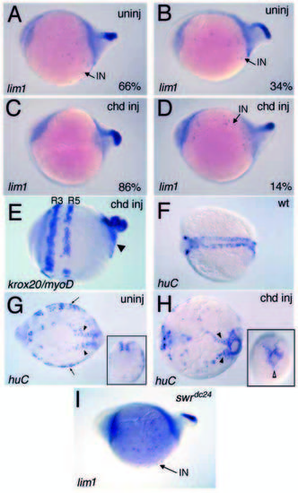
Expansion of lim1+ interneurons depends on Bmp activity. Lateral (A-E,I) and dorsal (F-H) views, anterior is to the left. In 6- to 7-somite uninjected swr/bmp2b homozygotes, 66% show a large expansion in lim1+ interneurons (A) and 34% exhibit a lesser or no expansion in interneurons (B). In the chordin-injected swr/bmp2b mutants, we see a shift to 86% with no or very few (<10) interneurons (C) and 14% that still exhibit normal or expanded numbers (D). Other head and trunk gene expression is unaffected as seen with krox20/myoD expression in a chordin-injected swr/bmp2b embryo (E). The arrowhead points to circular myoD expression in the somitic mesoderm. Expression of huC in 5- to 7-somite wild-type (F), uninjected (G) and chordin-injected (H) swr/bmp2b mutant embryos. (G) Arrows mark the lateral neural cells; (G,H) arrowheads indicate the medial trunk neural cells and insets are posterior views, dorsal to the top, showing a loss of lateral neural cells in ventral regions of injected mutants, while medial neural cells remain. (I) Expansion of lim1+ interneurons in swr/bmp2btdc24 presumptive null mutants. EXPRESSION / LABELING:
PHENOTYPE:
|

Examination of floor plate and motorneurons in dorsalized mutant embryos. shh expression in wild-type (A), swr/bmp2b (B), sbn/smad5 (C) and snh/bmp7 (D) mutant embryos at the 14-somite stage. The axis of the wild-type embryo (inset A) is straight, while the axis of the swr/bmp2b mutant (inset B) is curved. An intermediate dorsoventral spinal cord position of lim1+ cells in a cross-section of a 14-somite-stage wild-type embryo (E). Crosssections of 14-somite-stage embryos showing floor plate shh expression 1 cell wide in wild-type (F) and 2 to 5 cells wide in snh/bmp7 (G,J), swr/bmp2bsbn/smad5 (I). lim3 expression in the motorneurons of 7-somite-stage wild-type (K) and snh/bmp7 (L) mutant embryo. Expression of isl1 in the RB neurons and motorneurons in wild-type (M) and swr/bmp2b (N), where the motorneurons are apparent, but RB cells are absent. Anterior is to the top, dorsal to the right in A. Anterior is to the left, dorsal to the top in B-D. In K-N, dorsal views, anterior is to the left. FP, floor plate; P, pronephric region. EXPRESSION / LABELING:
PHENOTYPE:
|

Normal and expanded dorsal neural cell types in flh and ntl mutants, respectively. In 7-somite-stage embryos, rostral trunk regions of ntl mutants display normal numbers of tlx3- expressing RB precursors, while the caudalmost region, where shh expression is absent, exhibits an expansion in RB neurons (A,B, * marks the caudal region). (A′,B′) Enlargements of the posterior regions of A,B, showing the expanded RB cells in ntl mutants. In 10- somite-stage flh mutants, the RB cells and neural crest appear normal, even in regions where no shh-expressing midline cells are observed (C-F). (C′,D′) Enlargements of C,D showing weak shh expression in the tail bud of flh mutants. Caudal regions of ntl mutants display a mild expansion in the fkd6 neural crest domain at the 15-somitestage (G,H) and msxB expression in 7- somite-stage embryos (I,J). Arrowheads in G marks shh/fkd6 tail bud expression in wild type. Arrowheads in H indicate the expanded fkd6 expression domain in ntl mutants. (I′,J′) Enlargements of the posterior region of I,J, showing the loss of midline and tail bud shh expression in ntl mutants and expanded msxB expression. (A-J) Double in situ hybridizations including shh, except the insets in G and H where shh was omitted because tail bud shh expression obscures the fkd6 expression in flattened embryos. EXPRESSION / LABELING:
PHENOTYPE:
|

Absence of ventral motorneurons and presence of dorsal RB neurons in caudal segments of ntl mutants. The isl1- and lim3- expressing motorneurons are absent in caudal regions of ntl mutants, whereas the isl1-expressing RB neurons are present (A-D). Arrows mark the posterior extents of the expression domains. In caudal regions of ntl mutants, lim1-expressing interneurons (E,F) extend further posteriorly than the motorneurons (B,D), but do not extend as far posteriorly as the RB neurons (B). The lim1-expressing interneurons appear normal in rostral ntl segments, where shh expression is expanded in the floor plate (G,H). Anterior is to the left in A-F, dorsal to the top. Anterior is to the top in G,H, dorsal to the right. EXPRESSION / LABELING:
PHENOTYPE:
|