
|
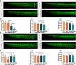
Fig. 5 NPs cause oxidative stress damage to embryonic CVP development. (A) Representative images of developmental defects in CVP after treatment with low-doses of H2O2 and/or NPs for 46 h. (B-D) The loop numbers (B), maximum ventral extension distance (C) and area (D) of embryonic CVP after NPs and/or H2O2 treatment (n = 30 embryos). (E) Representative images of embryonic CVP development after treatment with NAC and/or NPs for 46 h. (F-H) The loop numbers (F), maximum ventral extension distance (G), and area (H) of CVP in embryos treated with NPs and/or NAC (n = 30 embryos). Red asterisks, loop structure; white line, maximum ventral extension distance. Data are shown as mean ± SD. An asterisk above each bar indicates statistical significance (***P<0.001; ns: not significant). Scale bar, 100 μm.