
|
Image description by: Tanya Whitfield
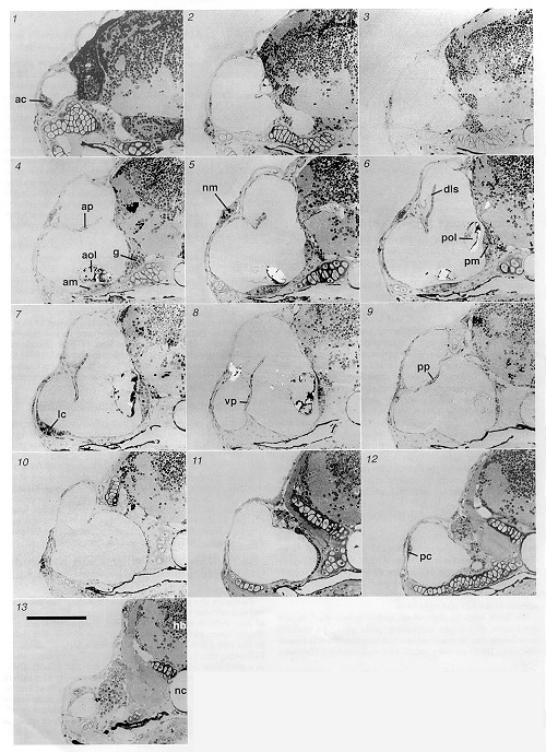
Anatomical structures shown: ear, sensory patches, semicircular canals, neuromast
Stage: stage series - 5 day
Genetic (background) strain: none given
Genotype: none given
Animal state: fixed
Labeling: toluidine blue stain
Description: Structure of a 5 day ear, illustrated by a series of 2 µm resin sections taken at 16 µm intervals. The first section is the most anterior. ac, anterior crista; am, anterior macula; aol, anterior otolith; ap, anterior pillar of semicircular canal; dls, dorsolateral septum (part of); g, ganglion; hb, hindbrain; lc, lateral crista; nc, notochord; nm, neuromast (lateral line organ); pc, posterior crista; pm, posteromedial macula; pol, posterior otolith; pp, posterior pillar of semicircular canal; vp, ventral pillar of semicircular canal. Scale bar, 100 µm. Reprinted by permission from the Journal of Comparative Neurology.
| Preparation | Image Form | View | Direction |
| LM-section | still | transverse | dorsal to top |