
-
- cancer (176)
- bacterial infectious disease (137)
- epilepsy (102)
- Parkinson's disease (90)
- breast cancer (70)
-

| Authors: | Amsterdam, A., Nissen, R.M., Sun, Z., Swindell, E., Farrington, S., and Hopkins, N. |
|---|---|
| Journal: | Proc. Natl. Acad. Sci. USA 2004 |
| Abstract: | We completed a large insertional mutagenesis screen in zebrafish to identify genes essential for embryonic and early larval development. We isolated 525 mutants, representing lesions in approximately 390 different genes, and we cloned the majority of these. Here we describe 315 mutants and the corresponding genes. Our data suggest that there are roughly 1,400 embryonic-essential genes in the fish. Thus, we have mutations in approximately 25% of these genes and have cloned approximately 22% of them. Re-screens of our collection to identify mutants with specific developmental defects suggest that approximately 50 genes are essential for the development of some individual organs or cell types. Seventy-two percent of the embryonic-essential fish genes have homologues in yeast, 93% have homologues in invertebrates (fly or worm), and 99% have homologues in human. Yeast and worm orthologues of genes that are essential for early zebrafish development have a strong tendency to be essential for viability in yeast and for embryonic development in the worm. Thus, the trait of being a genetically essential gene is conserved in evolution. This mutant collection should be a valuable resource for diverse studies of cell and developmental biology. |


| Authors: | Chen, J.-N., van Eeden, F.J.M., Warren, K.S., Chin, A., Nüsslein-Volhard, C., Haffter, P., and Fishman, M.C. |
|---|---|
| Journal: | Development 1997 |
| Abstract: | The first evident break in left-right symmetry of the primitive zebrafish heart tube is the shift in pattern of BMP4 expression from radially symmetric to left-predominant. The midline heart tube then 'jogs' to the left and subsequently loops to the right. We examined 279 mutations, affecting more than 200 genes, and found 21 mutations that perturb this process. Some cause BMP4 to remain radially symmetric. Others randomize the asymmetric BMP4 pattern. Retention of BMP4 symmetry is associated with failure to jog: right-predominance of the BMP4 pattern is associated with reversal of the direction of jogging and looping. Raising BMP4 diffusely throughout the heart, via sonic hedgehog injection, or the blocking of its action by injection of a dominant negative BMP4 receptor, prevent directional jogging or looping. The genes crucial to directing cardiac asymmetry include a subset of those needed for patterning the dorsoventral axis and for notochord and ventral spinal cord development. Thus, the pattern of cardiac BMP4 appears to be in the pathway by which the heart interprets lateralizing signals from the midline. |


| Authors: | Covassin, L., Amigo, J.D., Suzuki, K., Teplyuk, V., Straubhaar, J., and Lawson, N.D. |
|---|---|
| Journal: | Dev. Biol. 2006 |
| Abstract: | In this study, we utilize fluorescent activated cell sorting (FACS) of cells from transgenic zebrafish coupled with microarray analysis to globally analyze expression of cell type specific genes. We find that it is possible to isolate cell populations from Tg(fli1:egfp)(y1) zebrafish embryos that are enriched in vascular, hematopoietic and pharyngeal arch cell types. Microarray analysis of GFP(+) versus GFP(-) cells isolated from Tg(fli1:egfp)(y1) embryos identifies genes expressed in hematopoietic, vascular and pharyngeal arch tissue, consistent with the expression of the fli1:egfp transgene in these cell types. Comparison of expression profiles from GFP(+) cells isolated from embryos at two different time points reveals that genes expressed in different fli1(+) cell types display distinct temporal expression profiles. We also demonstrate the utility of this approach for gene discovery by identifying numerous previously uncharacterized genes that we find are expressed in fli1:egfp-positive cells, including new markers of blood, endothelial and pharyngeal arch cell types. In parallel, we have developed a database to allow easy access to both our microarray and in situ results. Our results demonstrate that this is a robust approach for identification of cell type specific genes as well as for global analysis of cell type specific gene expression in zebrafish embryos. |
| Authors: | Liu, D., Chu, H., Maves, L., Yan, Y.-L., Morcos, P.A., Postlethwait, J.H., and Westerfield, M. |
|---|---|
| Journal: | Development 2003 |
| Abstract: | The vertebrate inner ear develops from the otic placode, an ectodermal thickening that forms adjacent to the presumptive hindbrain. Previous studies have suggested that competent ectodermal cells respond to signals from adjacent tissues to form the placode. Members of the Fgf family of growth factors and the Dlx family of transcription factors have been implicated in this signal-response pathway. We show that compromising Fgf3 and Fgf8 signaling blocks ear development; only a few scattered otic cells form. Removal of dlx3b, dlx4b and sox9a genes together also blocks ear development, although a few residual cells form an otic epithelium. These cells fail to form if sox9b function is also blocked. Combined loss of Fgf signaling and the three transcription factor genes, dlx3b, dlx4b and sox9a, also completely eliminates all indications of otic cells. Expression of sox9a but not dlx3b, dlx4b or sox9b requires Fgf3 and Fgf8. Our results provide evidence for Fgf3- and Fgf8-dependent and -independent genetic pathways for otic specification and support the notion that Fgf3 and Fgf8 function to induce both the otic placode and the epithelial organization of the otic vesicle. |


| Authors: | Schilling, T.F., Piotrowski, T., Grandel, H., Brand, M., Heisenberg, C.P., Jiang, Y.J., Beuchle, D., Hammerschmidt, M., Kane, D.A., Mullins, M.C., van Eeden, F.J., Kelsh, R.N., Furutani-Seiki, M., Granato, M., Haffter, P., Odenthal, J., Warga, R.M., Trowe, T., and Nüsslein-Volhard, C. |
|---|---|
| Journal: | Development 1996 |
| Abstract: | Jaws and branchial arches together are a basic, segmented feature of the vertebrate head. Seven arches develop in the zebrafish embryo (Danio rerio), derived largely from neural crest cells that form the cartilaginous skeleton. In this and the following paper we describe the phenotypes of 109 arch mutants, focusing here on three classes that affect the posterior pharyngeal arches, including the hyoid and five gill- bearing arches. In lockjaw, the hyoid arch is strongly reduced and subsets of branchial arches do not develop. Mutants of a large second class, designated the flathead group, lack several adjacent branchial arches and their associated cartilages. Five alleles at the flathead locus all lead to larvae that lack arches 4-6. Among 34 other flathead group members complementation tests are incomplete, but at least six unique phenotypes can be distinguished. These all delete continuous stretches of adjacent branchial arches and unpaired cartilages in the ventral midline. Many show cell death in the midbrain, from which some neural crest precursors of the arches originate. lockjaw and a few mutants in the flathead group, including pistachio, affect both jaw cartilage and pigmentation, reflecting essential functions of these genes in at least two neural crest lineages. Mutants of a third class, including boxer, dackel and pincher, affect pectoral fins and axonal trajectories in the brain, as well as the arches. Their skeletal phenotypes suggest that they disrupt cartilage morphogenesis in all arches. Our results suggest that there are sets of genes that: (1) specify neural crest cells in groups of adjacent head segments, and (2) function in common genetic pathways in a variety of tissues including the brain, pectoral fins and pigment cells as well as pharyngeal arches. |


| Authors: | Alvarez, Y., Cederlund, M.L., Cottell, D.C., Bill, B.R., Ekker, S.C., Torres-Vazquez, J., Weinstein, B.M., Hyde, D.R., Vihtelic, T.S., and Kennedy, B.N. |
|---|---|
| Journal: | BMC Dev. Biol. 2007 |
| Abstract: | BACKGROUND: The retinal vasculature is a capillary network of blood vessels that nourishes the inner retina of most mammals. Developmental abnormalities or microvascular complications in the retinal vasculature result in severe human eye diseases that lead to blindness. To exploit the advantages of zebrafish for genetic, developmental and pharmacological studies of retinal vasculature, we characterised the intraocular vasculature in zebrafish. RESULTS: We show a detailed morphological and developmental analysis of the retinal blood supply in zebrafish. Similar to the transient hyaloid vasculature in mammalian embryos, vessels are first found attached to the zebrafish lens at 2.5 days post fertilisation. These vessels progressively lose contact with the lens and by 30 days post fertilisation adhere to the inner limiting membrane of the juvenile retina. Ultrastructure analysis shows these vessels to exhibit distinctive hallmarks of mammalian retinal vasculature. For example, smooth muscle actin-expressing pericytes are ensheathed by the basal lamina of the blood vessel, and vesicle vacuolar organelles (VVO), subcellular mediators of vessel-retinal nourishment, are present. Finally, we identify 9 genes with cell membrane, extracellular matrix and unknown identity that are necessary for zebrafish hyaloid and retinal vasculature development. CONCLUSIONS: Zebrafish have a retinal blood supply with a characteristic developmental and adult morphology. Abnormalities of these intraocular vessels are easily observed, enabling application of genetic and chemical approaches in zebrafish to identify molecular regulators of hyaloid and retinal vasculature in development and disease. |


| Authors: | Amacher, S.L., Draper, B.W., Summers, B.R., and Kimmel, C.B. |
|---|---|
| Journal: | Development 2002 |
| Abstract: | T-box genes encode transcriptional regulators that control many aspects of embryonic development. Here, we demonstrate that the mesodermally expressed zebrafish spadetail (spt)/VegT and no tail (ntl)/Brachyury T-box genes are semi-redundantly and cell-autonomously required for formation of all trunk and tail mesoderm. Despite the lack of posterior mesoderm in spt(-);ntl(-) embryos, dorsal-ventral neural tube patterning is relatively normal, with the notable exception that posterior medial floor plate is completely absent. This contrasts sharply with observations in single mutants, as mutations singly in ntl or spt enhance posterior medial floor plate development. We find that ntl function is required to repress medial floor plate and promote notochord fate in cells of the wild-type notochord domain and that spt and ntl together are required non cell-autonomously for medial floor plate formation, suggesting that an inducing signal present in wild-type mesoderm is lacking in spt(-);ntl(-) embryos. |


| Authors: | Aman, A., and Piotrowski, T. |
|---|---|
| Journal: | Dev. Cell 2008 |
| Abstract: | Collective cell migration is a hallmark of embryonic morphogenesis and cancer metastases. However, the molecular mechanisms regulating coordinated cell migration remain poorly understood. A genetic dissection of this problem is afforded by the migrating lateral line primordium of the zebrafish. We report that interactions between Wnt/beta-catenin and Fgf signaling maintain primordium polarity by differential regulation of gene expression in the leading versus the trailing zone. Wnt/beta-catenin signaling in leader cells informs coordinated migration via differential regulation of the two chemokine receptors, cxcr4b and cxcr7b. These findings uncover a molecular mechanism whereby a migrating tissue maintains stable, polarized gene expression domains despite periodic loss of whole groups of cells. Our findings also bear significance for cancer biology. Although the Fgf, Wnt/beta-catenin, and chemokine signaling pathways are well known to be involved in cancer progression, these studies provide in vivo evidence that these pathways are functionally linked. |


| Authors: | Anderson, R.M., Bosch, J.A., Goll, M.G., Hesselson, D., Dong, P.D., Shin, D., Chi, N.C., Shin, C.H., Schlegel, A., Halpern, M., and Stainier, D.Y. |
|---|---|
| Journal: | Dev. Biol. 2009 |
| Abstract: | Developmental mechanisms regulating gene expression and the stable acquisition of cell fate direct cytodifferentiation during organogenesis. Moreover, it is likely that such mechanisms could be exploited to repair or regenerate damaged organs. DNA methyltransferases (Dnmts) are enzymes critical for epigenetic regulation, and are used in concert with histone methylation and acetylation to regulate gene expression and maintain genomic integrity and chromosome structure. We carried out two forward genetic screens for regulators of endodermal organ development. In the first, we screened for altered morphology of developing digestive organs, while in the second we screed for the lack of terminally differentiated cell types in the pancreas and liver. From these screens, we identified two mutant alleles of zebrafish dnmt1. Both lesions are predicted to eliminate dnmt1 function; one is a missense mutation in the catalytic domain and the other is a nonsense mutation that eliminates the catalytic domain. In zebrafish dnmt1 mutants, the pancreas and liver form normally, but begin to degenerate after 84 hours post fertilization (hpf). Acinar cells are nearly abolished through apoptosis by 100 hpf, though neither DNA replication, nor entry into mitosis are halted in the absence of detectable Dnmt1. However, endocrine cells and ducts are largely spared. Surprisingly, dnmt1 mutants and dnmt1 morpholino-injected larvae show increased capacity for pancreatic beta cell regeneration in an inducible model of pancreatic beta cell ablation. Thus, our data suggest that Dnmt1 is dispensable for pancreatic duct or endocrine cell formation, but not for acinar cell survival. In addition, Dnmt1 may influence the differentiation of pancreatic beta cell progenitors or the reprogramming of cells toward the pancreatic beta cell fate. |


| Authors: | Ando, H., Kobayashi, M., Tsubokawa, T., Uyemura, K., Furuta, T., and Okamoto, H. |
|---|---|
| Journal: | Dev. Biol. 2005 |
| Abstract: | The telencephalon shows the greatest degree of size variation in the vertebrate brain. Understanding the genetic cascade that regulates telencephalon growth is crucial to our understanding of how evolution of the normal human brain has supported such a variation in size. Here, we present a simple and quick approach to analyze this cascade that combines caged-mRNA technology and the use of antisense morpholino oligonucleotides in zebrafish embryos. Lhx2, a LIM-homeodomain protein, and Six3s (Six3b and Six3a), another homeodomain proteins, show very similar expression patterns early in forebrain development, and these are known to be involved in the growth of this part of the brain. The telencephalon of six3b and six3a double morphant (six3 morphant) embryos is markedly reduced in size due to impaired cellular proliferation. Head-specific overexpression of Lhx2 by photoactivation of a caged-lhx2 mRNA completely rescued this size reduction, whereas similar head-specific activation of Six3b could not rescue the knockdown effect of lhx2. In the forebrain of medaka embryos, Six3 facilitates cellular proliferation by sequestration of Geminin from Cdt1, a key component in the assembly of the prereplication complex. Our results suggest that Lhx2 may mediate an alternative or parallel pathway for control of cellular proliferation in the developing forebrain via Six3. |


| Authors: | Arduini, B.L., Bosse, K.M., and Henion, P.D. |
|---|---|
| Journal: | Development 2009 |
| Abstract: | The neural crest generates multiple cell types during embryogenesis but the mechanisms regulating neural crest cell diversification are incompletely understood. Previous studies using mutant zebrafish indicated that foxd3 and tfap2a function early and differentially in the development of neural crest sublineages. Here, we show that the simultaneous loss of foxd3 and tfap2a function in zebrafish foxd3(zdf10);tfap2a(low) double mutant embryos globally prevents the specification of developmentally distinct neural crest sublineages. By contrast, neural crest induction occurs independently of foxd3 and tfap2a function. We show that the failure of neural crest cell diversification in double mutants is accompanied by the absence of neural crest sox10 and sox9a/b gene expression, and that forced expression of sox10 and sox9a/b differentially rescues neural crest sublineage specification and derivative differentiation. These results demonstrate the functional necessity for foxd3 and tfap2a for neural crest sublineage specification and that this requirement is mediated by the synergistic regulation of the expression of SoxE family genes. Our results identify a genetic regulatory pathway functionally discrete from the process of neural crest induction that is required for the initiation of neural crest cell diversification during embryonic development. |


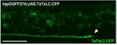
| Authors: | Asakawa, K., Suster, M.L., Mizusawa, K., Nagayoshi, S., Kotani, T., Urasaki, A., Kishimoto, Y., Hibi, M., and Kawakami, K. |
|---|---|
| Journal: | Proc. Natl. Acad. Sci. USA 2008 |
| Abstract: | Targeted gene expression is a powerful approach to study the function of genes and cells in vivo. In Drosophila, the P element-mediated Gal4-UAS method has been successfully used for this purpose. However, similar methods have not been established in vertebrates. Here we report the development of a targeted gene expression methodology in zebrafish based on the Tol2 transposable element and its application to the functional study of neural circuits. First, we developed gene trap and enhancer trap constructs carrying an engineered yeast Gal4 transcription activator (Gal4FF) and transgenic reporter fish carrying the GFP or the RFP gene downstream of the Gal4 recognition sequence (UAS) and showed that the Gal4FF can activate transcription through UAS in zebrafish. Second, by using this Gal4FF-UAS system, we performed large-scale screens and generated a large collection of fish lines that expressed Gal4FF in specific tissues, cells, and organs. Finally, we developed transgenic effector fish carrying the tetanus toxin light chain (TeTxLC) gene downstream of UAS, which is known to block synaptic transmission. We crossed the Gal4FF fish with the UAS:TeTxLC fish and analyzed double transgenic embryos for defects in touch response. From this analysis, we discovered that targeted expression of TeTxLC in distinct populations of neurons in the brain and the spinal cord caused distinct abnormalities in the touch response behavior. These studies illustrate that our Gal4FF gene trap and enhancer trap methods should be an important resource for genetic analysis of neuronal functions and behavior in vertebrates. |


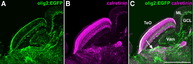
| Authors: | Bae, Y.K., Kani, S., Shimizu, T., Tanabe, K., Nojima, H., Kimura, Y., Higashijima, S.I., and Hibi, M. |
|---|---|
| Journal: | Dev. Biol. 2009 |
| Abstract: | The cerebellum is important for the integration of sensory perception and motor control, but its structure has mostly been studied in mammals. Here, we describe the cell types and neural tracts of the adult zebrafish cerebellum using molecular markers and transgenic lines. Cerebellar neurons are categorized to two major groups: GABAergic and glutamatergic neurons. The Purkinje cells, which are GABAergic neurons, express parvalbumin7, carbonic anhydrase8, and aldolase C like (zebrin II). The glutamatergic neurons are vglut1(+) granule cells and vglut2(high) cells, which receive Purkinje cell inputs; some vglut2(high) cells are eurydendroid cells, which are equivalent to the mammalian deep cerebellar nuclei. We found olig2(+) neurons in the adult cerebellum and ascertained that at least some of them are eurydendroid cells. We identified markers for climbing and mossy afferent fibers, efferent fibers, and parallel fibers from granule cells. Furthermore, we found that the cerebellum-like structures in the optic tectum and antero-dorsal hindbrain show similar Parvalbmin7 and Vglut1 expression profiles as the cerebellum. The differentiation of GABAergic and glutamatergic neurons begins 3 days post fertilization (dpf), and layers are first detectable 5 dpf. Using anti-Parvalbumin7 and Vglut1 antibodies to label Purkinje cells and granule cell axons, respectively, we screened for mutations affecting cerebellar neuronal development and the formation of neural tracts. Our data provide a platform for future studies of zebrafish cerebellar development. |


| Authors: | Balczerski, B., Matsutani, M., Castillo, P., Osborne, N., Stainier, D.Y., and Crump, J.G. |
|---|---|
| Journal: | Dev. Biol. 2012 |
| Abstract: | Development of the head skeleton involves reciprocal interactions between cranial neural crest cells (CNCCs) and the surrounding pharyngeal endoderm and ectoderm. Whereas elegant experiments in avians have shown a prominent role for the endoderm in facial skeleton development, the relative functions of the endoderm in growth versus regional identity of skeletal precursors have remained unclear. Here we describe novel craniofacial defects in zebrafish harboring mutations in the Sphingosine-1-phospate (S1P) type 2 receptor (s1pr2) or the S1P transporter Spinster 2 (spns2), and we show that S1P signaling functions in the endoderm for the proper growth and positioning of the jaw skeleton. Surprisingly, analysis of s1pr2 and spns2 mutants, as well as sox32 mutants that completely lack endoderm, reveals that the dorsal–ventral (DV) patterning of jaw skeletal precursors is largely unaffected even in the absence of endoderm. Instead, we observe reductions in the ectodermal expression of Fibroblast growth factor 8a (Fgf8a), and transgenic misexpression of Shha restores fgf8a expression and partially rescues the growth and differentiation of jaw skeletal precursors. Hence, we propose that the S1P-dependent anterior foregut endoderm functions primarily through Shh to regulate the growth but not DV patterning of zebrafish jaw precursors. |


| Authors: | Becker, T.S., Burgess, S.M., Amsterdam, A.H., Allende, M.L., and Hopkins, N. |
|---|---|
| Journal: | Development 1998 |
| Abstract: | Not really finished (nrf), a larval-lethal mutation in zebrafish generated by retroviral insertion, causes specific retinal defects. Analysis of mutant retinae reveals an extensive loss of photoreceptors and their precursors around the onset of visual function. These neurons undergo apoptosis during differentiation, affecting all classes of photoreceptors, suggesting an essential function of nrf for the development of all types of photoreceptors. In the mutant, some photoreceptors escape cell death, are functional and, as judged by opsin expression, belong to at least three classes of cones and one class of rods. The protein encoded by nrf is a close homologue of human Nuclear Respiratory Factor 1 and avian Initiation Binding Repressor, transcriptional regulators binding the upstream consensus sequence RCGCRYGCGY. At 24 hours of development, prior to neuronal differentiation, nrf is expressed ubiquitously throughout the developing retina and central nervous system. At 48 hours of development, expression of nrf is detected in the ganglion cell layer, in the neurons of the inner nuclear layer, and in the optic nerve and optic tracts, and, at 72 hours of development, is no longer detectable by in situ hybridization. Mutants contain no detectable nrf mRNA and die within 2 weeks postfertilization as larvae with reduced brain size. On the basis of its similarity with NRF-1 and IBR, nrf is likely involved in transcriptional regulation of multiple target genes, including those that encode mitochondrial proteins, growth factor receptors and other transcription factors. This demonstrates the power of insertional mutagenesis as a means for characterizing novel genes necessary for vertebrate retinal development. |


| Authors: | Bedell, V.M., Person, A.D., Larson, J.D., McLoon, A., Balciunas, D., Clark, K.J., Neff, K.I., Nelson, K.E., Bill, B.R., Schimmenti, L.A., Beiraghi, S., and Ekker, S.C. |
|---|---|
| Journal: | Development 2012 |
| Abstract: | The Homeobox (Hox) and Paired box (Pax) gene families are key determinants of animal body plans and organ structure. In particular, they function within regulatory networks that control organogenesis. How these conserved genes elicit differences in organ form and function in response to evolutionary pressures is incompletely understood. We molecularly and functionally characterized one member of an evolutionarily dynamic gene family, plac8 onzin related protein 1 (ponzr1), in the zebrafish. ponzr1 mRNA is expressed early in the developing kidney and pharyngeal arches. Using ponzr1-targeting morpholinos, we show that ponzr1 is required for formation of the glomerulus. Loss of ponzr1 results in a nonfunctional glomerulus but retention of a functional pronephros, an arrangement similar to the aglomerular kidneys found in a subset of marine fish. ponzr1 is integrated into the pax2a pathway, with ponzr1 expression requiring pax2a gene function, and proper pax2a expression requiring normal ponzr1 expression. In addition to pronephric function, ponzr1 is required for pharyngeal arch formation. We functionally demonstrate that ponzr1 can act as a transcription factor or co-factor, providing the first molecular mode of action for this newly described gene family. Together, this work provides experimental evidence of an additional mechanism that incorporates evolutionarily dynamic, lineage-specific gene families into conserved regulatory gene networks to create functional organ diversity. |


| Authors: | Behra, M., Gallardo, V.E., Bradsher, J., Torrado, A., Elkahloun, A., Idol, J., Sheehy, J., Zonies, S., Xu, L., Shaw, K.M., Satou, C., Higashijima, S.I., Weinstein, B.M., and Burgess, S.M. |
|---|---|
| Journal: | BMC Dev. Biol. 2012 |
| Abstract: | BackgroundBecause of the structural and molecular similarities between the two systems, the lateral line, a fish and amphibian specific sensory organ, has been widely used in zebrafish as a model to study the development/biology of neuroepithelia of the inner ear. Both organs have hair cells, which are the mechanoreceptor cells, and supporting cells providing other functions to the epithelium. In most vertebrates (excluding mammals), supporting cells comprise a pool of progenitors that replace damaged or dead hair cells. However, the lack of regenerative capacity in mammals is the single leading cause for acquired hearing disorders in humans. ResultsIn an effort to understand the regenerative process of hair cells in fish, we characterized and cloned an egfp transgenic stable fish line that trapped tnks1bp1, a highly conserved gene that has been implicated in the maintenance of telomeres' length. We then used this Tg(tnks1bp1:EGFP) line in a FACsorting strategy combined with microarrays to identify new molecular markers for supporting cells. ConclusionsWe present a Tg(tnks1bp1:EGFP) stable transgenic line, which we used to establish a transcriptional profile of supporting cells in the zebrafish lateral line. Therefore we are providing a new set of markers specific for supporting cells as well as candidates for functional analysis of this important cell type. This will prove to be a valuable tool for the study of regeneration in the lateral line of zebrafish in particular and for regeneration of neuroepithelia in general. |


| Authors: | Beis, D., Bartman, T., Jin, S.W., Scott, I.C., D'Amico, L.A., Ober, E.A., Verkade, H., Frantsve, J., Field, H.A., Wehman, A., Baier, H., Tallafuss, A., Bally-Cuif, L., Chen, J.N., Stainier, D.Y., and Jungblut, B. |
|---|---|
| Journal: | Development 2005 |
| Abstract: | Defects in cardiac valve morphogenesis and septation of the heart chambers constitute some of the most common human congenital abnormalities. Some of these defects originate from errors in atrioventricular (AV) endocardial cushion development. Although this process is being extensively studied in mouse and chick, the zebrafish system presents several advantages over these models, including the ability to carry out forward genetic screens and study vertebrate gene function at the single cell level. In this paper, we analyze the cellular and subcellular architecture of the zebrafish heart during stages of AV cushion and valve development and gain an unprecedented level of resolution into this process. We find that endocardial cells in the AV canal differentiate morphologically before the onset of epithelial to mesenchymal transformation, thereby defining a previously unappreciated step during AV valve formation. We use a combination of novel transgenic lines and fluorescent immunohistochemistry to analyze further the role of various genetic (Notch and Calcineurin signaling) and epigenetic (heart function) pathways in this process. In addition, from a large-scale forward genetic screen we identified 55 mutants, defining 48 different genes, that exhibit defects in discrete stages of AV cushion development. This collection of mutants provides a unique set of tools to further our understanding of the genetic basis of cell behavior and differentiation during AV valve development. |


| Authors: | Ben, J., Elworthy, S., Ng, A.S., van Eeden, F., and Ingham, P.W. |
|---|---|
| Journal: | Development 2011 |
| Abstract: | Using zinc-finger nuclease-mediated mutagenesis, we have generated mutant alleles of the zebrafish orthologue of the chicken talpid3 (ta3) gene, which encodes a centrosomal protein that is essential for ciliogenesis. Animals homozygous for these mutant alleles complete embryogenesis normally, but manifest a cystic kidney phenotype during the early larval stages and die within a month of hatching. Elimination of maternally derived Ta3 activity by germline replacement resulted in embryonic lethality of ta3 homozygotes. The phenotype of such maternal and zygotic (MZta3) mutant zebrafish showed strong similarities to that of chick ta3 mutants: absence of primary and motile cilia as well as aberrant Hedgehog (Hh) signalling, the latter manifest by the expanded domains of engrailed and ptc1 expression in the somites, reduction of nkx2.2 expression in the neural tube, symmetric pectoral fins, cyclopic eyes and an ectopic lens. GFP-tagged Gli2a localised to the basal bodies in the absence of the primary cilia and western blot analysis showed that Gli2a protein is aberrantly processed in MZta3 embryos. Zygotic expression of ta3 largely rescued the effects of maternal depletion, but the motile cilia of Kupffer’s vesicle remained aberrant, resulting in laterality defects. Our findings underline the importance of the primary cilium for Hh signaling in zebrafish and reveal the conservation of Ta3 function during vertebrate evolution. |


| Authors: | Bergeron, S.A., Tyurina, O.V., Miller, E., Bagas, A., and Karlstrom, R.O. |
|---|---|
| Journal: | Development 2011 |
| Abstract: | The transmembrane protein Brother of Cdo (Boc) has been implicated in Shh-mediated commissural axon guidance, and can both positively and negatively regulate Hedgehog (Hh) target gene transcription, however, little is known about in vivo requirements for Boc during vertebrate embryogenesis. The zebrafish umleitung (uml(ty54)) mutant was identified by defects in retinotectal axon projections. Here, we show that the uml locus encodes Boc and that Boc function is cell-autonomously required for Hh-mediated neural patterning. Our phenotypic analysis suggests that Boc is required as a positive regulator of Hh signaling in the spinal cord, hypothalamus, pituitary, somites and upper jaw, but that Boc might negatively regulate Hh signals in the lower jaw. This study reveals a role for Boc in ventral CNS cells that receive high levels of Hh and uncovers previously unknown roles for Boc in vertebrate embryogenesis. |