
- Title
-
Dissecting hematopoiesis and disease using the zebrafish
- Authors
- Amatruda, J.F. and Zon, L.I.
- Source
- Full text @ Dev. Biol.
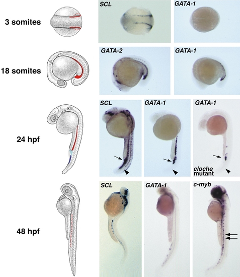
The expression pattern of hematopoietic factors during zebrafish embryogenesis. Whole-embryo in situ hybridization analysis showing the localization of hematopoietic factors in zebrafish embryos at 3 somites, 18 somites, 24 h, or 48 h postfertilization (hpf). In the schematic panels (left), colored regions mark the anatomic areas involved in embryonic hematopoiesis. Early markers such as SCL are expressed in two stripes of lateral plate mesoderm by the 3-somite stage. GATA-1 expression, not detectable at this stage, begins at approximately 5 somites. Between 18 somites and 24 hpf the stripes converge and fuse to form the ICM (red, anterior ICM; light blue, posterior ICM; dark blue, ventral tail region). Unlike SCL, which is expressed throughout the ICM and in the ventral tail, GATA-1 expression is limited to the anterior and posterior ICM (arrows, posterior ICM; arrowheads, ventral tail putative hematopoietic region). In cloche mutant embryos, GATA-1 persists only in the posterior ICM. By 48 hpf, SCL and GATA-1 expression in blood is decreased, though SCL is highly expressed in the brain. c-myb is expressed in the ventral wall of the dorsal aorta (double arrow). 3 somites, dorsal view; 18 somites, anterior is to the left, dorsal is up; 24 and 48 hpf, anterior is up, dorsal is to the right. (Courtesy of C. Kimmel, M. A. Thompson, D. G. Ransom, and E. C. Liao.) |

Hematopoiesis in zebrafish embryos at 4 days postfertilization. Expression of SCL, c-myb, and rag-1 at 4 dpf. SCL and c-myb are expressed in the ventral tail region (arrows) as well as the nervous system. rag-1 expression is confined to the thymus (arrowheads). (Courtesy of E. C. Liao and N. Trede.) |

Positive and negative cis-acting elements regulating hematopoietic expression of GATA-1. Transgenic zebrafish expressing green fluorescent protein from the GATA-1 promoter were generated by microinjection of GATA-1–GFP constructs and selection of transgenic founder lines. 18-somite-stage progeny are shown. In (A), the transgene contains the full-length GATA-1 promoter . GFP expression is confined to hematopoietic progenitors. In (B), deletion of a negative cis-acting element leads to ectopic GFP expression in notochord (arrow); hematopoietic expression is not affected. (Courtesy of Dr. Shuo Lin.) |
Reprinted from Developmental Biology, 216(1), Amatruda, J.F. and Zon, L.I., Dissecting hematopoiesis and disease using the zebrafish, 1-15, Copyright (1999) with permission from Elsevier. Full text @ Dev. Biol.