
- Title
-
her1, a zebrafish pair-rule like gene, acts downstream of notch signalling to control somite development
- Authors
- Takke, C. and Campos-Ortega, J.A.
- Source
- Full text @ Development
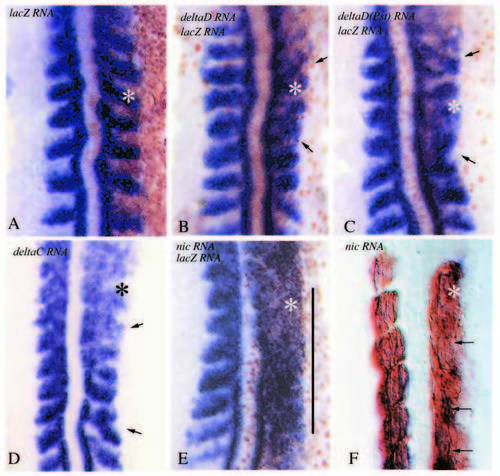
(A-G) Flat preparations of embryos injected with various RNAs. Asterisks label the affected side. (A) 10-somite stage control embryo injected with lacZ RNA alone. (B,C) 10- somite-stage embryos injected with full-length deltaD (B) and with deltaD(Pst) (C) and lacZ RNA. Both embryos have been stained for MyoD (blue, in situ hybridization) and β-galactosidase (brown, antibody staining) expression. Somites are irregularly shaped on the affected side, arrows point to somite fusions. (D) 10-somitestage embryo injected with full-length deltaC RNA and stained by in situ hybridization for MyoD expression. Somitic defects are similar to those in B and C. Arrows point to somite fusions. (E) 10-somite-stage embryo injected with notch1a-intra (nic) mRNA and lacZ RNA and stained for MyoD (blue, in situ hybridization) and β-galactosidase (brown, antibody staining) expression. Notice the poor somitic organization on the affected side, MyoD expression is diffuse and no somite boundaries can be distinguished in the territory labelled by the vertical line. (F) 22- h-stage embryo that had been injected with notch1a-intra (nic) mRNA and lacZ RNA, and stained for myosin heavy chain. Notice that the outlines of the somites appear blurred and muscle fibres are not packed into somitic groups. The arrows point to individual muscle fibres extending through regions in which somite borders should have developed |

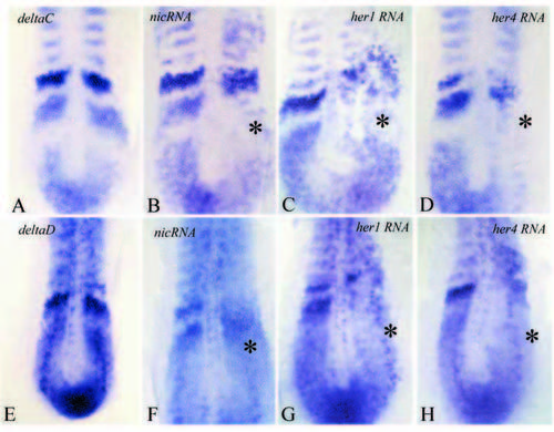
Flat preparations of 8- to 10- somite-stage embryos injected with notch1a (nic) (B,G), deltaD (C,H) and deltaC (D,I) and deltaD(Pst) RNA (E,K). (A,F) Normal embryos. All embryos have been processed for in situ hybridization with two probes, either MyoD (red) and her1 (blue, A-E) or MyoD (red) and her4 (blue, F-K). Asterisks in B-E and GK label the affected side; arrows point to her1 and her4 domains (blue staining). (B) On the affected side of notch1a-intra (nic) RNA injected embryos, her1 is upregulated within the presomitic mesoderm, but not in the immediate neighbourhood of the somitic territory, and the characteristic her1 stripes cannot be distinguished. Notice that MyoD expression is also unpatterned in the somitic mesoderm. Following nic RNA injection, her4 expression is also upregulated in the presomitic mesoderm on the injected side (notice that there are more blue stained cells on the injected side), MyoD expression in the somitic territory is diffuse (G). Following injection of deltaD (C,H), deltaC (D,I) or deltaD(Pst) RNA (E,K), somitic defects are visible as manifested by the MyoD expression (asterisks). However, her1 and her4 expression are essentially normal. The her1 stripes are clearly separated from each other and the presomitic expression of her4 is as weak as in normal embryos (arrows point to blue cells, compare with F). The diffuse blue colour on the injected side of the embryos in H, I and K is due to activation of her4 transcription in the neural plate (Takke et al., 1999). |

Somitic defects following injection of her1 (A,B), her4 (C) or her1 and her4 (D) RNA. (A,C,D) In situ hybridizations with MyoD; (B) an anti-myosin staining. A-C have also been processed with an anti-β-galactosidase antibody. Asterisks label the affected side, arrows in A and C point to partially fused somites. The severity of defects in A-C is comparable to that in embryos injected with delta variants (see Fig. 1). Note that somite borders have formed and muscle fibres insert at the borders of neighbouring myotomes (arrows). However, somitic organization is less clearcut, in fact practically absent, in embryos injected simultaneously with her1 and her4 RNA (vertical bar in D). |
Expression patterns of deltaC (A-D) and deltaD (E-H) following injection of nic (B,F), her1 (C,G) or her4 (D,H) RNA. (A,E) Normal embryos. Asterisks label the affected side. Notice considerable patterning defects and reduction of the amount of transcript on the injected side. Refer to text for further details. |
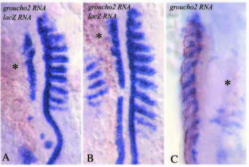
Flat preparations of embryos injected with groucho2 and lacZ mRNAs and stained for MyoD (blue, in situ hybridization) and β- galactosidase (brown, antibody staining) expression (A,B), and for pax-9 (blue, in situ hybridization) and myosin heavy chain (brown, antibody staining) expression (C). Asterisks label the affected side. Mesodermal differentiation is strongly reduced. |