
- Title
-
The smad5 mutation somitabun blocks Bmp2b signaling during early dorsoventral patterning of the zebrafish embryo
- Authors
- Hild, M., Dick, A., Rauch, G.J., Meier, A., Bouwmeester, T., Haffter, P., and Hammerschmidt, M.
- Source
- Full text @ Development
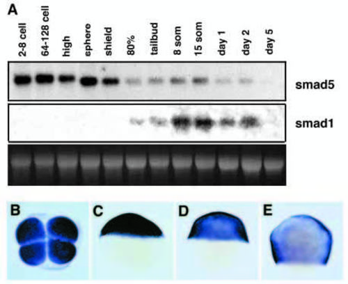
Expression of smad5 and smad1. (A) Developmental northern analysis with 10 μg of total RNA of the indicated stages, hybridized with a smad5- (upper panel) and a smad1- (middle panel) specific probe. Lower panel shows ethidium bromide staining of 28S rRNA as loading control. (B-H) Whole-mount in situ hybridization using a smad5-specific probe. (B) 4-cell stage, animal view. (C) Sphere stage, lateral view. (D) Shield stage, lateral view, dorsal right. (F) 80% epiboly, lateral view, dorsal right. |

Later phenotypes of sbntc24 mutant embryos, 36 hpf, lateral view. (A) Wild type; (B) dominant maternal effect of sbn, causing strong C4 dorsalization. (C) Dominant zygotic effect of sbn, causing weak C1 dorsalization (arrow to ventral tail fin deficiency). (D) sbn,swr double heterozygote from a cross of a swr heterozygous female and a sbn heterozygous male, characterized by a loss of the ventral tail fin and a wound-up tail (C3, arrow), indicating a significantly stronger dorsalization than in C. (E-H) The maternal effect of sbn in the background of different sbn genotypes, 48 hpf, lateral view; (E-G) Embryos from a cross of two sbn heterozygous parents; (H) embryo from a cross of a sbn homozygous female with a heterozygous male. Both crosses led to offspring with 100% C4 dorsalization. The maternal and zygotic genotypes of individual embryos are indicated. The zygotic genotype was determined by tetra-PCR after photography. |
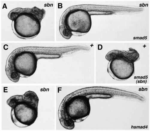
Rescue and phenocopy of sbntc24 mutant phenotype. The genotype of embryos is indicated in the upper right corner, the injected RNA in the lower right corner; 36 hpf, lateral view. (A,C,E) Uninjected controls; (B,D,F) injected embryos. (A,B,E,F) Rescue of sbn phenotype upon injection of wild-type smad5 mRNA (A,B) or human SMAD4 mRNA (E,F) into embryos from a cross of two sbn heterozygous parents. (C,D) Phenocopy of sbn phenotype upon injection of smad5 mRNA, bearing the sbn mutation, into wild-type embryos. |
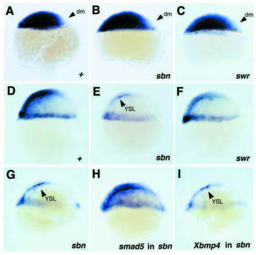
Expression of bmp2b in sbn and swr mutant embryos (A-F) and after injection of smad5 or Xbmp4 mRNA in sbn mutants (G-I). All embryos are shown in a lateral view, dorsal side right. (A,D) Wild-type siblings of swr mutants; (B,E) sbn mutants from cross of sbn heterozygous female with wild-type male. Embryos were fixed and stained in parallel to the swr and wild-type embryos shown; (C,F) swr mutants. (A-C) Sphere stage; presumptive dorsal mesoderm (dm) devoid of bmp2b staining is marked with arrowheads. (D-F) Shield stage; the sbn mutant (E) displays severely reduced bmp2b staining, and the swr mutant (F) slightly reduced bmp2b staining in the blastoderm; the staining in the yolk syncytial layer (YSL, arrowhead in E) appears normal. (G-I) sbn mutant embryos, shield stage, after injection of smad5 (H; 50 pg/embryo) or Xbmp4 (I; 4 pg/embryo) mRNA. Similar results as in I were obtained for approx. 100 embryos in each of three independent injection experiments. Of the Xbmp4-injected sbn sibling embryos that were raised to day 2, 53% (n=45) showed a strong morphological response, appearing wild-type or ventralized (see Fig. 6). |

Rescue of ventral cell fates in sbn mutant cells by exogenous bmp4 mRNA or DNA (A-D) and wild-type environment (E-H); 36 hpf, lateral view. (A,B) Injection of 1 pg Xbmp4 mRNA into a sbn homozygous mutant embryo from a cross of two homozygous mutant parents. The injected embryo displays a normalization of the sbn mutant phenotype from C4 (A) to C1 (B, arrow indicates absence of ventral tail fin). (C,D) Injection of 50 pg pCSKA-bmp4 DNA into an embryo from a cross of two sbn heterozygous parents. The sbn mutant phenotype is converted from a strong dorsalization (C4, panel C) to a weak ventralization (V1, panel D; see slight duplication of the ventral tail fin (arrow) and the enlarged blood island (arrowhead). (E-H) Chimeric embryos with fluorescein-labeled cells from embryos of a sbn/+ x sbn/+ cross (E,G) or a sbn/sbn x sbn/sbn cross (F,H) transplanted into wild-type recipients. Donor cells give rise to blood (E,F) and ventral tail fin (G,H) (see also Table 2). |