
- Title
-
Epidermal expression of apolipoprotein E gene during fin and scale development and fin regeneration in zebrafish
- Authors
- Monnot, M.J., Babin, P.J., Poleo, G., Andre, M., Laforest, L., Ballagny, C., and Akimenko, M.A.
- Source
- Full text @ Dev. Dyn.

Figure 1. Expression of apoE during pectoral fin morphogenesis. A-D: ApoE transcripts are detected starting at the onset of pectoral fin bud outgrowth and become restricted to the apical margin of the bud. Lateral view of A: 31-hr; B: 35-hr ; C: 3-day ; D: 7-day, 3 mm, zebrafish embryos and larva, anterior to the left. The red arrow in A indicates the pectoral fin bud emerging from the yolk sac. Note that apoE is strongly expressed in the yolk sac throughout development. E: pectoral fin of a 50-day, 10.3 mm, embryo. During larval stage, apoE is expressed in the epidermis of the distal part of the fin (black arrow), along the anterior margin of the fin (red arrow), and in the inter-ray tissues (white arrow). F: pectoral fin of adult fish, 32 mm. ApoE is still expressed in the pattern as during larval stage, albeit at a much lower level. y, yolk sac. Scale bar = 100 m. |
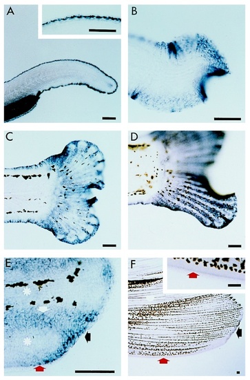
Figure 2. Expression of apoE during caudal fin morphogenesis. Lateral view, anterior to the left. A: 35-hr embryo. ApoE is expressed in the epidermis of the unique median fin fold around the posterior part of the embryo. Inset is a high magnification showing that there is, however, no expression in the most distal layer of cells. B: 7-days, 3 mm, larva. ApoE transcripts become more widely distributed in the epidermis of the tail region. C-E: During larval stages, apoE is expressed in the epidermis surrounding the developing fin rays. C: 21-day, 5.6 mm; D: 24-day, 6.3 mm ; E: 40-day, 10 mm larvae. E: High magnification of two developing fin rays (asterisks) shows that apoE is expressed in the epidermis of the distal part of the fin (black arrow), along the margin of the fin but not in the most external epidermal cell layer (red arrow), and in the inter-ray tissue (white arrow in E). F: ventral lobe of the caudal fin of an adult fish, 33 mm. ApoE is still expressed in the epidermis in the same pattern as during larval development (arrows) but at a much lower level. The inset is a high magnification of the lateral fin margin showing that there is no expression in the most distal epidermal cell layer. Scale bar = 100 m. |
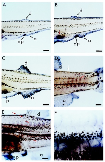
Figure 3. Expression of apoE during dorsal, anal, and pelvic fins morphogenesis and during scale development. Lateral view, anterior to the left of A: 19-day, 4.8 mm B: 22-day, 5.7 mm ; C: 24-day, 6.3 mm; D: 30-day, 8.5 mm; E: 40-day, 10 mm; F: 50-day, 10.8 mm larvae. A-C: Since the embryonic stage, apoE is always expressed in the median epithelial fin fold. During the larval stages, apoE is expressed in the anal (a) and dorsal (d) fins which emerge from the fin fold, and in the pelvic fin buds (red arrow in B and (p) in C). Note that apoE is also strongly expressed in the anal papilla (ap). D: Later, apoE expression disappears in the epidermis where the median fin fold has completely regressed (white arrow), while the fins are still expressing apoE. ApoE transcripts are also detected as the scale papillae form starting in the caudal part of the trunk (red arrow). E-F: As the scale papillae develop, the domain of apoE-expressing cells becomes restricted to the posterior periphery of the scale (E : red arrow in the ventral part of the larva, and F). a, anal fin; ap, anal papilla; c, caudal fin; d, dorsal fin; p, pelvic fin. Scale bar = 200 m. EXPRESSION / LABELING:
|

Figure 4. ApoE expression is enhanced during regeneration of the pectoral fin. ApoE expression was determined A:18 hr; B: 24 hr; C: 3 days; D: 4 days; E: 6 days; F: 14 days after amputation. The plane of amputation is indicated by the thick red arrow. A-E: apoE is strongly expressed in the wound epidermis at the apex of each fin ray during the first 4 to 6 days of regeneration. F: 14 days after amputation, apoE expression has decreased to levels comparable to control non-amputated pectoral fin. A thin red arrow indicates a differentiating lepidotrichia in D,E and a newly formed bifurcation in F. ae, apical epidermis; b, blastema; we, wound epidermis. Scale bar = 200 m. EXPRESSION / LABELING:
|

Figure 5. ApoE expression during regeneration of the caudal fin. ApoE expression was determined A: 12 hr; B: 16 hr; C,D: 24 hr; F,H:2 days; E: 3 days; G,J: 4 days; L: 6 days; I: 14 days; K: 30 days after amputation. (A,C,E,G,I,K) whole-mount in situ hybridization. (B,D,F,J,L) proximo-distal and (H) transverse sections after in situ hybridization. The plane of amputation is indicated by a thick red arrow in A-G. A,C: apoE expression was observed as early as 12 hr after amputation at the apex of each truncated lepidotrichia. E,G: a strong expression was detected in the apical epidermis of each fin ray during the first 4 to 6 days of regeneration. I: 14 days after amputation, apoE expression has considerably decreased. K: 30 days after amputation, apoE expression levels are comparable to control non-amputated fin. A thin red arrow indicates a formed bifurcation in I and apoE expression along the lateral side of the fin in K. B: frozen section along the proximo-distal axis of a fin 16 hr after amputation shows that apoE is expressed in the epidermal cells covering the wound as well as in putative macrophages (thin red arrow). F: putative macrophages expressing apoE are also observed 2 days after amputation. D: 24 hr after amputation, apoE is expressed in the basal layer of the epidermis. F,H,J,L: Throughout the regeneration process, apoE is abundantly expressed in the basal layer of the apical epidermis and to a lesser extend in the intermediate cell layers. ApoE transcripts are never detected in the most superficial epidermal layer. H: transverse section shows that apoE is expressed at a low level in the basal epidermal layer of the inter-ray tissue. ae, apical epidermis; b, blastema; be, basal layer of the epidermis; l, lepidotrichia ; we, wound epidermis. A,C,E,G,I,K scale bar = 200 m ; B,D,F,H,J,L scale bar = 20 m. EXPRESSION / LABELING:
|