
- Title
-
Delta-Notch signaling and the patterning of sensory cell differentiation in the zebrafish ear: evidence from the mind bomb mutant
- Authors
- Haddon, C., Jiang, Y.-J., Smithers, L., and Lewis, J.
- Source
- Full text @ Development
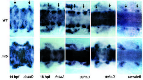
Expression of deltaA, deltaB, deltaD and serrateB in wild-type embryos (WT, top row) and mib mutant embryos (bottom row) at placode (14 hpf) and early otocyst (18 hpf) stages of ear development, shown by whole-mount in situ hybridization. The photographs are dorsal views of the ear rudiments and the hindbrain region between them; arrows indicate sites of delta and serrate gene expression at the anterior and posterior ends of the otic placode/otocyst. Anterior is to the left. Note the intensified and expanded expression of the genes at these sites in mib mutants – most obvious for deltaD and serrateB, but evident also for deltaA and deltaB. The band of deltaD-expressing cells very strikingly seen in the 14 hpf mib specimen extending between the anterior and posterior poles of the otocyst may correspond to the region from which neuroblasts will later delaminate. EXPRESSION / LABELING:
PHENOTYPE:
|

Tracing cell fate by labelling with lysinated rhodamine dextran (LRDX). In this example, a single cell injected with LRDX at the anterior site of delta gene expression in an early otocyst (18 hpf; A) ends up in the anteroventral macula, where it gives rise to a hair cell plus a supporting cell (identified by their shape and location in the epithelium; B and C). The pictures are superpositions of fluorescence and transmission-optics images taken with a confocal microscope in the living embryo at the time of injection and 24 and 48 hours afterwards. The specimen shown is one of approximately 80 that were used to build up a fate map (Haddon, 1997). In other specimens, labelled cells at the same original site end up in the same location. ao, anterior otolith; po, posterior otolith; am, anterior macula; hc, hair cell. |

(A,B) Expression of deltaA, deltaB and deltaD in wild-type ears at 30 and 42 hpf, shown by in situ hybridisation on cryosections cut transverse to the main body axis. Arrows indicate scattered cells expressing delta genes in the thickened ventral wall of the otocyst. Beneath this epithelium, in the 30 hpf deltaA and deltaB sections, one can see delta gene expression in cells of the statoacoustic ganglion. (C) Expression of deltaB in a cell in the hair-cell layer of a sensory patch (the anteroventral macula) at 42 hpf; such deltaB-expressing hair cells are few and far between, because expression of the gene fades rapidly as a hair cell matures. (D) Persistent and extensive expression of serrateB, seen at 4 days in the hair-cell layer of the posteromedial and anteroventral maculae (arrows). |

Expression of deltaA, B and D in the neural plate in wild-type and mib embryos at 12 hpf (6-7 somites); a part of the trunk region is shown in dorsal view, as in Fig. 1. The mutation causes increased delta expression but also increased numbers of primary neurons, showing that the mutant cells are refractory to the inhibitory effect of Delta-Notch signalling. EXPRESSION / LABELING:
PHENOTYPE:
|
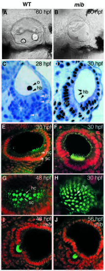
Wild-type and mib ears compared. (A,B) Lateral views of live embryos (Nomarski optics) at 60 hpf. Note that the ears are smaller and deformed in mib and that the otoliths are drastically reduced, reflecting a deficit in the sensory patches. (C-D) Semi-thin Araldite sections stained with toluidine blue, showing the altered pattern of cell differentiation. (E-H) Confocal images of whole-mount specimens stained with fluorescent phalloidin (green) to reveal actin-rich hair bundles and with 7AAD (red nuclear staining) as a counterstain. (E,F) Optical sections passing transversely through sensory patches; (G,H) En-face views of sensory patches. Supporting cells are missing in mib, while hair cells are present in great excess. (I,J) Stages in the extrusion of hair cells from the otic epithelium in mib; staining is as in E-H. The hair cells eventually degenerate and disappear. hc, hair cell; sc, supporting cell; o, otolith; hb, hair bundle; n, neuron. |
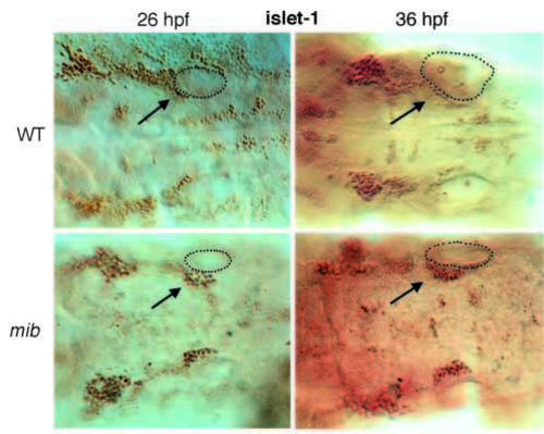
Dorsal view of ears plus hindbrain at 26 and 36 hpf, stained with islet-1 antibody using HRP detection, showing the statoacoustic ganglion (arrows) containing many more neurons in mib than in wild type. The outline of the right ear is indicated by a dotted line. The statoacoustic ganglion at early stages forms a continuous mass with the anterior lateral line ganglion and the VIIth cranial nerve ganglion. As a rough guide, however, one can take the statoacoustic ganglion cells to be those that lie at and posterior to the anterior end of the otocyst; we followed this rule in making the counts described in the text. |

Unillustrated author statements PHENOTYPE:
|