
- Title
-
Molecular identification of spadetail: regulation of zebrafish trunk and tail mesoderm formation by T-box genes
- Authors
- Griffin, K.J.P., Amacher, S.L., Kimmel, C.B., and Kimelman, D.
- Source
- Full text @ Development
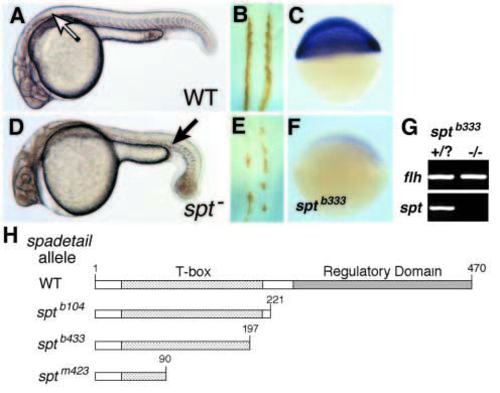
Appearance of (A) wild-type (WT) and (D) spt- live embryos at 24 hours postfertilization (h.p.f.; anterior, left). In both panels, arrow marks the first visible somite. Chevron-shaped somites are visible in the trunk and tail of WT embryos, but are absent from the trunk of spt- embryos. In spt- embryos, presumptive trunk mesodermal progenitors migrate abnormally and form a mass of cells at the tail tip (Ho and Kane, 1990). (B,E) Dorsal views of 24 h.p.f. embryos stained with an antibody to mero-myosin (anterior, uppermost). Bilateral rows of expression are visible either side of the notochord in the WT embryo (B) but staining in the spt- embryo (E) is significantly reduced and is patchy. (C,F) Embryos at 50% epiboly obtained from intercrosses of sptb333/+ adults hybridized with spt probe (blue stain). 75% of the embryos (C) express spt, whereas 25% of the embryos from such a cross, which presumably represent the mutant embryos (F), had no detectable spt expression, even after a prolonged colour reaction. (G) Bulk-segregant analysis of sptb333 genomic DNA by PCR. As a control, floating head (flh) was amplified from pooled DNA isolated from phenotypically WT and spt- embryos, whereas the spt candidate gene could not be amplified from the mutant DNA. (H) Schematic maps of WT and truncated Spt proteins; the putative regulatory domain is inferred from studies of the Brachyury protein (Kispert et al., 1995a). Numbers above the coding regions refer to amino acid position. See Methods for details of the molecular lesions. EXPRESSION / LABELING:
PHENOTYPE:
|

Expression of spt in WT and spt- embryos and regulation by FGF signaling. (A) spt expression, first detected between sphere and dome stages, is ubiquitous in the late blastula embryo (40% epiboly, 5 h.p.f., lateral view). (B) By the early gastrula stage (shield, 6 h.p.f., dorsal view), spt expression is specific to the mesoderm and, in an animal pole view (C), is found throughout all regions of the germ ring (d, dorsal). (E) spt expression refines during gastrulation and by 60% epiboly (7 h.p.f., dorsal view), spt is not expressed in the notochord progenitors (n), whereas lateral and ventral germ ring and head mesoderm (arrow) express spt. (F,G) Vegetal views of spt expression in late-gastrula embryos (80% epiboly, 8.3 h.p.f., dorsal uppermost). (F) Expression of a dominant-negative FGF receptor (dnFGFR), which is mosaicly distributed (Griffin et al., 1995), causes loss of spt expression in large areas of the germ ring; compare with WT in G. (I) At the 4-somite stage (11.5 h.p.f., anterior to left) expression is localized to the hatching gland progenitors (arrow), the tail bud and the segmental plate mesoderm. (J,K) In the tail bud, spt is expressed in epiblast cells at the site of involution (arrowhead in J, * in K), and in the segmental plate (S); spt is also expressed in adaxial cells (arrows; Devoto et al., 1996). (J, 8 somites, 13 h.p.f., lateral view, anterior to left; K, 4 somites, 11.5 h.p.f., posterior view, dorsal uppermost). (D,H,L) spt expression in spt- embryos. (D) In early gastrula spt- embryos (shield, 6 h.p.f., animal pole view), spt expression is weak but has normal spatial distribution (d, dorsal); compare with C. (H) During gastrulation (80% epiboly, 8.3 h.p.f., vegetal view), spt expression is not maintained in hypoblast or dorsolateral epiblast, but is weakly expressed on the ventral side; compare with G. (L) In early-somite stage spt- embryos (4 somite, 11.5 h.p.f., posterior view, dorsal uppermost), spt is expressed in the tail bud (*) but not in the segmental plate mesoderm (compare with K). EXPRESSION / LABELING:
|

Overlapping and complementary expression of T-box genes in the early mesoderm. (A-C) Sections through lateral germ ring at midgastrulation (8.5 h.p.f.); arrows mark limits of expression in epiblast (e) and hypoblast (h). (A) spt and (B) tbx6 are expressed in epiblast cells closest to the margin, and broadly in the hypoblast. (C) ntl expression is expressed only near the margin in both the epiblast and hypoblast. (D-F) Posterior views of 4-6-somite embryos (12 h.p.f., dorsal uppermost). (D) spt and (E) tbx6 are co-expressed in the region of the tail bud epiblast (*) where involution occurs (Kanki and Ho, 1997) and in the underlying hypoblast and segmental plate (S), but only spt is prominently expressed in adaxial cells (arrows in D). (F) ntl is expressed more broadly in the tail bud epiblast (*); ntl expression in the hypoblast is restricted to the notochord (arrow). EXPRESSION / LABELING:
|
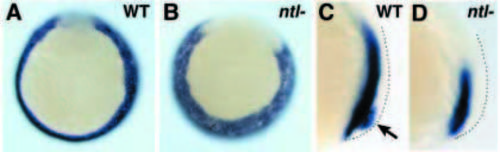
Dynamic regulation of spt expression in ntl- embryos. (A,B) Mid gastrula (7.5 h.p.f., vegetal view). spt is expressed at normal levels in the ntl mutant embryo. Although the spatial distribution of spt expression is slightly different in ntl- embryos, the broader region of non-expressing cells represent dorsal mesoderm as they express the axial mesoderm marker floating head (data not shown). (C,D) 8-somite stage (13 h.p.f., lateral view, anterior is left). spt expression in the tail bud epiblast (arrow) is readily detected in WT embryos (C), but this expression domain is not seen in ntl- embryos at a similar stage (D). The domain of spt expression in involuted mesoderm is only partially reduced in the mutant embryo. The increased thickness of the epiblast in ntl- embryos is already apparent at this stage (dots mark outer surface of epiblast). EXPRESSION / LABELING:
|

spt function regulates tbx6 expression in trunk but not tail mesoderm. (A) In WT embryos at the early gastrula stage (shield, 6 h.p.f., animal pole view; d, dorsal) tbx6 expression is restricted to ventral mesoderm. During gastrulation (B; 80% epiboly, 8.3 h.p.f., vegetal view) tbx6 expression is up-regulated in the dorsolateral mesoderm, and during segmentation (C,D) tbx6 is extensively expressed in tail bud epiblast (*) and segmental plate (C, 2-4 somites, 11 h.p.f.; D, 7-8 somites, 13 h.p.f.; posterior view, dorsal uppermost). In spt- embryos, tbx6 expression is initially weak (E), remains restricted to the ventral side during gastrulation (F) and is barely detectable in the segmental plate at 2-4 somites (G). However, at 7-8 somites (H) tbx6 has a relatively normal distribution compared to WT, and is easily detected in segmental plate mesoderm (arrows) in addition to the tail bud (*). EXPRESSION / LABELING:
|

Synergistic regulation of spt and tbx6 by spt and ntl. Expression of tbx6 (A-D; bud, 10 h.p.f.) and spt (E-H; 95% epiboly, 9.5 h.p.f.). In ntl- embryos expression of tbx6 (B) and spt (F) are similar to WT (A,E) except for broadening of the non-expressing notochord domain (Melby et al., 1997). In addition, spt is no longer prominently expressed by adaxial cells. In spt- embryos, expression of both tbx6 (C) and spt (G) are very weak or absent from hypoblast cells but remain detectable in the tail bud epiblast (*). In spt-;ntl- embryos, expression of tbx6 (D) is entirely undetectable, and spt (H) is only barely detected (arrow). EXPRESSION / LABELING:
|