
- Title
-
Genes controlling and mediating locomotion behavior of the zebrafish embryo and larva
- Authors
- Granato, M., van Eeden, F.J., Schach, U., Trowe, T., Brand, M., Furutani-Seiki, M., Haffter, P., Hammerschmidt, M., Heisenberg, C.P., Jiang, Y.J., Kane, D.A., Kelsh, R.N., Mullins, M.C., Odenthal, J., and Nüsslein-Volhard, C.
- Source
- Full text @ Development

Comparison of muscle differentiation between wild-type (A,C,E,G), slo (B,D,F,J), fro (I) and fub (H) embryos using polarized light (A-F) or trunk muscle sections (G-J). At 30 hours birefringency is reduced in slo embryos (B) compared to wild-type embryos (A). At later stages (96 hours) muscle birefringency is detectable in somitic muscle and in jaw muscle of wild-type larvae (C, E), while in slo mutants (D, F) birefringency is completely absent in somitic and jaw muscles. The arrows in E delineate the jaw muscles. (G) Sagitallateral section through 36 hours wild-type somitic muscle tissue showing muscle fibers (arrows) and elongated nuclei (arrowhead). In fub mutants muscle fibers are absent; however, the nuclei are elongated (arrowhead) and resemble those of wild type (H). In contrast, nuclei in fro (I) and slo (J) are round (arrowhead). (J) Occasionally muscle fibers can be detected in slo somitic tissue (arrows). |

Comparison of muscle birefingency between wild type and muscle mutants. Muscle fibers are affected in tur (B), slp (D), dus (E) and buf (G) mutants compared to wild type (A,C,F). Optical section through lateral myotomes in a 72-hour wild-type embryo (A) shows muscle fibers in the dorsal half of the somites. In tur mutants, similar to all mutants in the phenotypic groups A2 and A3, birefringency appears reduced or disorganized (B). In slp mutants about 20% fewer muscle fibers are visible (D) and in dus mutants (E) only residual fibers can be seen (arrowheads), compared to wild-type embryos at 60 hours (C). In wild-type muscle tissue (F) segmental organization of the sarcomeres is visible; the arrowheads indicate three individual units. In buf mutants sarcomeric organization is perturbed (G). Sarcomeres appear less evenly spaced and reduced in size. PHENOTYPE:
|

Loss of birefringency and lesions in the somites become apparent in somite degeneration mutants. In sap (B) and ruz (C) mutants birefringency is decreased around 96 hours, compared to wild type (A), and lesions become visible in the somitic muscle tissue. (D) Lateral view of a wild-type larva around 96 hours; the somitic segments are separated by distinct boundries. (E,F) In sap and sof mutants degeneration affects a few individual somitic segments (black arrowheads indicate somite boundaries). The ventral part of the affected somite (E) appears normal (between white arrowheads). (G) In ruz mutants degeneration is affecting all somites. PHENOTYPE:
|

Motorneuron development is affected in unp mutants. (A) The three primary motorneurons in the ventral half of the spinal cord (sc), CaP, MiP and RoP, occupy unique positions along the antero-posterior axis and extend stereotypic, axonal projections. (B) The antibody znp-1 stains the CaP axons in the ventral and the MiP axons in the dorsal somites of wild-type embryos. (C) In unp mutants MiP axons appear to elongate normally toward dorsal, while axons extending ventrally have abnormal morphology. They form multiple branches (arrowheads) and do not extend as far as in wildtype embryos. Whether these aberrant axons originate from CaP somatas is not clear. CaP, caudal primary motorneuron; MiP, middle primary motorneuron; RoP, rostral primary motorneuron; d, dorsal; v, ventral; sc, spinal cord; n, notochord; hm, horizontal myoseptum. |

Retinotecal projection phenotype in maomao mutants these axons defasciculate prematurely in the anterior half of the tectum (B,D). a, anterior tectum; p, posterior tectum. PHENOTYPE:
|

Wild-type and ‘accordion’ behavior at around 30 hours. Wildtype embryos (A-F) and beo mutant embryos (G-L) were liberated from their chorions and their behavior monitored. Wild-type embryos move their tail from one side of the body to the other (A-F). In contrast, ‘accordion’ group mutants contract along their body axis (G-L). A line behind the yolk extension is added as a constant reference. The lengths of the horizontal arrows reflect the contraction along the antero-posterior axis of the embryo. The vertical arrows point to the region in the anterior notochord that becomes compressed during the contraction (H,I). PHENOTYPE:
|

Notochord lesions in ‘accordion’ group mutants. Lateral view on a 60-hour live wild type (A) and acc mutant (B). (A) Shows a wild-type notochord, demarcated by two arrows. In acc embryos (B), local lesions in the notochord become visible (arrowheads); next to this lesion the notochord appears unaffected (arrows). PHENOTYPE:
|
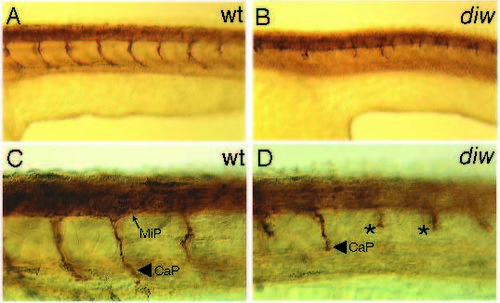
Axonal outgrowth of primary motorneurons are affected in diw mutants. Antibody labeling with the znp-1 antibody stains axonal projections of primary motorneurons in 30-hour wild type (A,C) and residual axons in diw mutant embryos (B,D). (A) Lateral view of a wild-type embryo stained with znp-1. Per segment, one CaP axon is visible. In diw mutants these axons are shorter (B). (C) Higher magnification of the same wild-type embryo as in A. The MiP and the CaP axons are labeled. (D) Higher magnification of the same diw embryo as in B. Short, CaP-like axons are stained (asterisk). No axons resembling dorsally projecting MiP axons are labeled. |

Behavioral phenotype of the twitch twice group. Wild type (A-F) and spc mutant larvae (G-L) locomotion behavior were recorded at 120 hours. Single frame sequences illustrate alternating tail movements in wild-type larvae (A-F). Mutant spc larvae do not alternate their tail movements, but bend their tail consecutively towards the same side (G-L). PHENOTYPE:
|
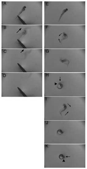
tnt larvae show exaggerated alternating tail movements. Single frames from a video showing a fast response upon tactile stimulus of a wild-type larva at around 60 hours (A-D). An arrow indicates the direction in which the larva is moving. tnt mutant larvae show alternating, but hyperactive tail movements (E-K). One such cycle is shown, where the tail touches the head (H) on one side, then the embryo moves its tail to the other side and again touches the head (K). In H and K: head, arrow; tail, arrowhead. PHENOTYPE:
|

Retinotectal projection defect in nev mutants. Retinotectal projections of dye-labeled nasodorsal axons in wild-type (A) and nev (B) larvae at 120 hours. In wild type the retinotectal projections terminate in the posteriodorsal optic tectum (small arrowhead in A), while in nev mutants (B) they terminate on both posteriodorsal (small arrowhead) and posterioventral positions (arrow). The large arrowheads in A and B indicate the position of the dye injection. d, dorsal tectum. PHENOTYPE:
|

ZFIN is incorporating published figure images and captions as part of an ongoing project. Figures from some publications have not yet been curated, or are not available for display because of copyright restrictions. PHENOTYPE:
|
|
ZFIN is incorporating published figure images and captions as part of an ongoing project. Figures from some publications have not yet been curated, or are not available for display because of copyright restrictions. PHENOTYPE:
|
|
ZFIN is incorporating published figure images and captions as part of an ongoing project. Figures from some publications have not yet been curated, or are not available for display because of copyright restrictions. PHENOTYPE:
|
|
Unillustrated author statements |