
- Title
-
Mutations affecting xanthophore pigmentation in the zebrafish, Danio rerio
- Authors
- Odenthal, J., Rossnagel, K., Haffter, P., Kelsh, R.N., Vogelsang, E., Brand, M., van Eeden, F.J., Furutani-Seiki, M., Granato, M., Hammerschmidt, M., Heisenberg, C.P., Jiang, Y.J., Kane, D.A., Mullins, M.C., and Nüsslein-Volhard, C.
- Source
- Full text @ Development

ZFIN is incorporating published figure images and captions as part of an ongoing project. Figures from some publications have not yet been curated, or are not available for display because of copyright restrictions. PHENOTYPE:
|

Adult fish that are heterozygous or homozygous for salz or pfeffer show pigment pattern defects. (A) Wild-type, (B) heterozygous pfetg17 and (C) homozygous saltt254a fish in a lateral view. PHENOTYPE:
|
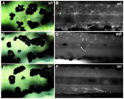
Xanthophore pigment is reduced in edison and brie. Day 6 larva, dorsal view of the head region posterior to the eyes, viewed with nomarski optics (A,C,E) and lateral view of ammonia-induced fluorescence in the trunk under UV light epi-illumination (B,D,F). Wild type (A,B) and homozygous mutant larvae of edito255b (C), editc245c (D), and britj226a (E,F). M, melanophores; X and arrow, xanthophores. PHENOTYPE:
|
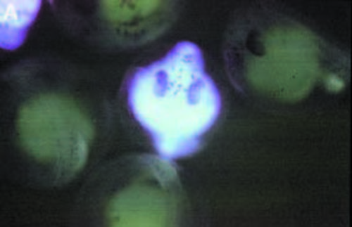
Mutant edison embryos show strong autofluorescence under UV-light illumination. Embryos at the pharyngula period derived from two heterozygous carriers for editk232. Homozygous mutant edi embryos show strong blue fluorescence in the entire embryo, whereas wild-type siblings show only background levels. PHENOTYPE:
|
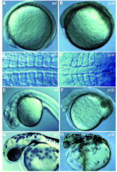
Embryos derived from homozygous mutant yobty44d females show a maternal effect phenotype. Lateral view of (A) a wild-type embryo and (B) an embryo derived from a yob homozygous female at the 5-somite stage, showing the reduced length of the body. (C) Dorsal view of a 10-somite stage wild-type embryo and (D) a 10- somite stage embryo derived from a yob homozygous female showing the wider axis. Lateral view of a wild-type embryo (E) and an embryo derived from a yob homozygous female (F) at the pharyngula period, showing the head and tail defects. Lateral view of a wild-type embryo (G) and an embryo derived from a yob homozygous female (H) at the hatching period. PHENOTYPE:
|
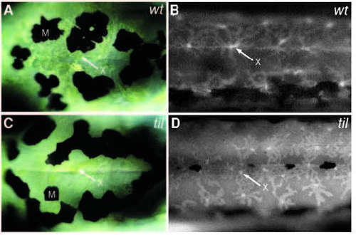
In tilsit mutants xanthophore pigment appears condensed whereas the cells appear more spread out. Dorsal view of day-6 larva in the head region posterior to the eyes, viewed with Nomarski optics (A,C), and lateral view of ammonia-induced fluorescence in the trunk under UV-light epi-illumination (B,D). (A,B) Wild type and (C,D) homozygous mutant larva of tilty130b. M, melanophores; X, and arrow, xanthophores. PHENOTYPE:
|