
- Title
-
Coordinate embryonic expression of three zebrafish engrailed genes
- Authors
- Ekker, M., Wegner, J., Akimenko, M.A., and Westerfield, M.
- Source
- Full text @ Development

Cells at the junction between the midbrain and hindbrain express all three engrailedgenes. Parasagittal sections through the heads of 12 h (A-C) and 32 h (E-G) embryos and 22 h (D) and 32 h (H) whole-mount embryos were hybridized with probes for the eng1 (A,E), eng2 (8,F) or eng3 (C,D,G,H) genes. A cartoon of the engrailed expression domains at the midbrain/hindbrain junction is shown in (I). The domain expressing only eng3 is shown in blue, the domain expressing both eng2 and eng3 is shown in red, and the domain expressing all three is shown in green. Anterior is oriented to the left and dorsal to the top in this and subsequent figures. AC and E-G are double exposures with bright field (blue) and clarkfield (red) illumination. The red signal in the lower part of I A is due to the yolk, rather than specific hybridization, as shown in control sections without hybridization. Arrows in H indicate hindbrain cells expressing eng3 (see Fig. 8). Scale bar, 80 μm in A-D, 50 μm in E-G and I, 100 μm in H. EXPRESSION / LABELING:
|

The muscle pioneers express the eng1 and eng2 genes. Parasagittal sections through the trunk myotomes of 16 h (A,B) and 24 h (C,D) embryos and 36 h whole-mount embryos (E) were hybridized with probes for the eng1 (A,C,E) or eng2 (B,D) genes. The locations of the muscle pioneers are indicated by the arrows. The red signals in the lower part of D and the brown signals in E are due to pigment cells, rather than specific hybridization, as shown in unhybridized control sections. Scale bar, 50 μm in A,B,D and 35 μm in C and E. EXPRESSION / LABELING:
|
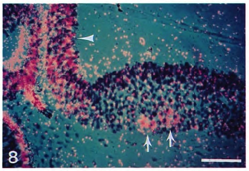
Cells in the first three hindbrain rhombomeres express the eng3 gene. A parasagittal section through the hindbrain of a 32 h embryo was hybridized with a probe for the eng3 gene. Small clusters of cells (arrows) in the ventral parts of the second and third rhomobomeres hybridize. A cluster of cells in the first rhombomere (contained in a different section, not shown, and as illustrated by the whole-mount embryo in Fig. 6 H) also hybridized. The hybridization at the junction between the midbrain and hindbrain is also apparent (arrowhead). The signal in the lower left is caused by the pigment epithelium of the retina and is not due to specific hybridization as shown in unhybridized control sections. Scale bar, 50 μm. EXPRESSION / LABELING:
|

Jaw muscle precursors express the eng2 and eng3 genes. Parasagittal sections of 32 h embryos were hybridized with probes for the eng1 (A), eng2 (8), or eng3(C) genes. Precursors of the jaw muscles (arrows) are indicated. The signal around the eye in each panel is caused by the pigment epithelium of the retina rather than to specific hybridization as shown in unhybridized control sections. Scale bar, 50 μm. EXPRESSION / LABELING:
|

Ectodermal cells in the pectoral fins express the eng1 gene. Parasagittal sections through the distal portion of the pectoral fin buds of 32 h embryos were hybridized with probes for the engt (top) , eng2(middle) or eng3 (bottom) genes. The lateral ectoderm in the ventral part of the bud (arrows) hybridized with only the eng1 probe. The red signals in the lower part of Band C are due to the yolk, rather than specific hybridization, as shown in unhybridized control sections. Scale bar, 50 μm. EXPRESSION / LABELING:
|