
- Title
-
Expression of snail2, a second member of the zebrafish Snail family, in cephalic mesendoderm and presumptive neural crest of wild-type and spadetail mutant embryos
- Authors
- Thisse, C., Thisse, B., and Postlethwait, J.H.
- Source
- Full text @ Dev. Biol.
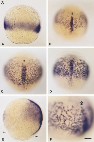
Distribution of snail2 RNA during gastrulation. Transcripts from the snail2 gene were revealed by whole-mount in situ hybridization. (A) Optical section of an embryo at 65% epiboly oriented with its animal pole up. snail2 RNA appears in involuted mesendoderm. (B) Dorsal view of an embryo at 70% epiboly. The dorsal axial mesoderm appears as a nearly continuous strip of snail2 labeling about 3 cells wide coursing through the network of snail2-expressing cells. Presumptive hatching gland cells (asterisk) that form the anterior part of the prechordal plate do not express snail2. (C) Embryo at 80% epiboly in a dorsal–anterior view or (D) in dorsal–posterior view. The dorsal axis staining has become broader and extends beyond the stained web. (E) Optical section of an embryo at 90% epiboly. Dorsal, right, and animal pole, top. The axial snail2 expression domain extends posteriorly in the notochordal territory and reaches the margin (indicated by arrowheads). (F) High magnification of the left side of an embryo at 90% epiboly in the cephalic region. Cells labeled with snail2 probe form branching rows from 10 to 20 cells long. Scale bars, 25 μm (A–E); 40 μm (F). |
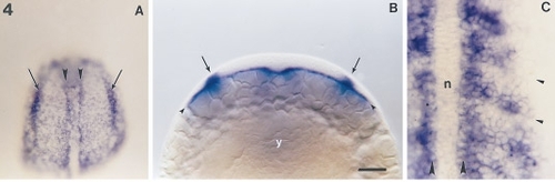
Expression pattern of snail2 at the end of gastrulation and beginning of somitogenesis. Embryos were dissected from the yolk in A and C. (A) Embryo with three somites in dorsal view, anterior to the top. Two rows of strongly labeled snail2-expressing cells, called cephalic adaxial cells (arrowheads), are continuous with adaxial cells in the trunk bordering the unlabeled notochord. The cephalic adaxial cells define the border of the prechordal plate. Laterally, presumptive neural crest cells (arrows) begin to accumulate snail2 transcript. (B) An optical section in the presumptive head of an embryo at the same stage showing the mesendoderm (arrowheads), a very thin layer of cells expressing snail2, lying adjacent to the yolk cell (y). Medial to the lateral borders of the stained cephalic mesoderm, snail2 transcripts are revealed in presumptive neural crest cells (arrows). (C) Dorsal view of the trunk of a seven-somite embryo. The notochord (n) no longer contains snail2 transcript. Adaxial cells (large arrowheads) are strongly labeled as well as cells of the posterior part of the somites. Somitic furrows are indicated by small arrowheads. Scale bars, 25 μm (A), 40 μm (B), and 100 μm (C). |
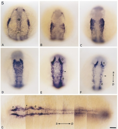
snail2 expression during somitogenesis. (A–F) dorsal view of embryos at (A) 3-somite stage, (B) 5-somite stage, (C) 7-somite stage, (D) 9-somite stage, (E) 12-somite stage, (F) 14-somite stage. Mesendodermal labeling is still strong in the prechordal plate (asterisk) at the 3-somite stage but has disappeared from the notochord (n), producing a sharp border between these two structures (two dashes). Mesendodermal staining fades as more somites form (B–G). snail2 transcripts are observed in cells lining the neural plate, in particular in neural crest cells. snail2 expression is detected when convergence of neural crest occurs toward the midline (A–D), and then as they migrate ventrally or anteriorly away from the neural keel (E–F), around structures like optic vesicles (o), ears (e), or trigeminal ganglia (arrowhead). (G) Photomontage of an embryo at the 14-somite stage in dorsal view. Anteriorly, snail2-expressing cells partially surround the optic vesicles (o), trigeminal ganglia (arrowhead), and the ear (e). In the anterior part of the embryo, neural crest cells have already begun their migration. In the trunk snail2-expressing cells are dorsal in the neural keel. This behavior illustrates the wave of anteroposterior differentiation of neural crest cells that occurs during somitogenesis. Anteroposterior (a, p) axis is indicated and is vertical from A to F and horizontal in G. Scale bar, 25 μm. |

snail2 expression pattern during late somitogenesis in the tail and in the otic vesicle region. (A) Expression pattern of snail2 in the posterior trunk and tail bud of an embryo at the 16-somite stage. On the dorsal side of the embryo (toward the top of the figure) snail2 staining in the neural crest extends 12 somites anteriorly from the tail bud (arrowheads). Ventrally, the staining is very strong in mesoderm. (B) An optical section through the tail region of the same embryo viewed at the level of the dashes in A (dorsal, top). snail2 transcripts appear in neural crests dorsally in the neural rod (between arrowheads) and in the medial and ventral part of the somite. Less intense staining occurs in muscle pioneer precursors located lateral to the notochord (n). (C) Tail of a 24-hr-old embryo. snail2 expression appears at the position of neural crest in 3 to 4 of the most recently formed somites (between arrowheads). (D) Optical section of a 24-hr-old embryo at the level of the otic vesicle (e) and (E) lateral view of the same embryo. snail2 transcripts accumulate just ventral and anterior to the otic vesicle. dorsoventral (d–v) and anteroposterior (a–p) axis. Scale bars, 25 μm (A), 40 μm (B–E). |
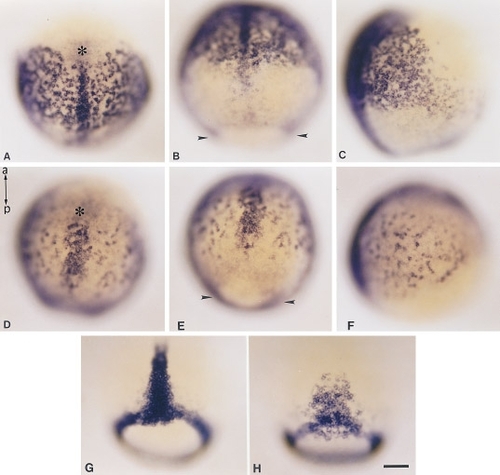
Expression pattern of snail2 and no tail in wild-type and spadetail (sptb104) mutant embryos at gastrulation. (A–C) Wild-type and (D–F) sibling spadetail embryos at 90% epiboly. (A and D) Dorsoanterior view; (B and E) dorsoposterior view; (C and F) right side views. An asterisk marks the presumptive hatching gland domain and an arrowhead indicates the marginal zone. spadetail embryos have fewer snail2-expressing cells laterally than wild type. snail2-expressing cells in the axis occupy an irregular, loose, and broader domain compared to wild type. The snail2 expression domain is also closer to the margin. This shows that a spadetail embryo is shorter than wild type and that the axial domain of snail2 expression is enlarged at gastrulation. This broadening of axial mesoderm was confirmed by examining the distribution of no tail RNA, a notochord marker (Schulte-Merker et al., 1994) in wild-type (G) and spadetail mutant (H) embryos at 80% epiboly. Anteroposterior (a–p). Scale bar, 25 μm. |

Expression pattern of snail2 in wild-type and spadetail (sptb104) mutant embryos at the end of gastrulation and beginning of somitogenesis. (A, C, E) Wild-type embryos and (B, D, F) sibling spadetail mutant embryos in dorsoposterior views. (A, B) Embryos at the end of gastrulation; (C, D) embryos at bud stage; (E, F) embryos at the two-somite stage. Labeled adaxial cells (ad) lining the notochord territory are observed only in wild-type embryos. The posterior limits of the snail2 expression domain in the lateral and paraxial cephalic mesendoderm are indicated by open arrows and are closer to the margin (arrowheads in A and B) or the tail bud (tb) in spadetail mutant embryos than in wild type. Neural crest labeling (indicated by arrows in E and F) appears identical in wild-type and mutant embryos. Scale bar, 25 μm. |
Reprinted from Developmental Biology, 172, Thisse, C., Thisse, B., and Postlethwait, J.H., Expression of snail2, a second member of the zebrafish Snail family, in cephalic mesendoderm and presumptive neural crest of wild-type and spadetail mutant embryos, 86-99, Copyright (1995) with permission from Elsevier. Full text @ Dev. Biol.