
- Title
-
The protein product of the zebrafish homologue of the mouse T gene is expressed in nuclei of the germ ring and the notochord of the early embryo
- Authors
- Schulte-Merker, S., Ho, R.K., Herrmann, B.G., and Nüsslein-Volhard, C.
- Source
- Full text @ Development

Temporal expression pattern of Zf-T. 10 μg of total RNA of the indicated stages were blotted. Staging was done according to Westerfield (1989). A single transcript of about 2.5 kb was detected upon probing with the entire insert of pBSCT-ZFc1. The left panel shows methylene blue staining of ribosomal RNA of the same blot. |
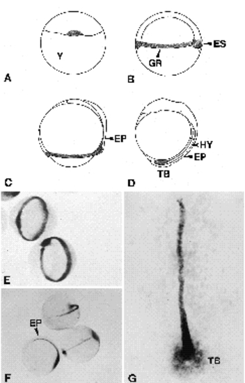
Distribution of Zf-T mRNA in early embryos. (A-D) Schematic drawings of embryos, indicating (shaded) the distribution of Zf-T mRNA as revealed by whole-mount in situ hybridizations. Embryos are oriented with their animal pole up, and with their dorsal side to the right (in A, the dorsal-ventral polarity is not discernable yet). (A) Sphere stage, 4.0 hours. The blastomeres sit on top of the yolk (Y), epiboly is just about to start. (B) Embryonic shield stage, 6.0 hours. Epiboly has led to a spreading of cells over the yolk mass, which has undergone a change in shape. Involution causes a thickening of the embryo around its equator. Dorsal convergence leads to a thickening of the germ ring (GR) at the dorsal side, the embryonic shield (ES). (C) 80% epiboly, 8.0 hours. The embryo is two-layered: cells that have involuted constitute the hypoblast (HY; future endo- and mesoderm), cells that have not involuted constitute the epiblast (EP). (D) 2 somite-stage, 11.0 hours. Epiboly has finished, but gastrulation continues in the tailbud (TB) which is going to extend over the next couple of hours to make up the posterior half of the body. (E-G) Whole-mount in situ hybridizations using antisense RNA probes to reveal spatial expression of Zf-T. (E) Embryonic shield stage. Cells in the region of the germ ring express high levels of Zf-T RNA. Note the thickening at the dorsal side, the embryonic shield (ES). (F) Tailbud stage. Zf-T RNA is expressed in the presumptive notochord and in the tailbud (TB), but not in cells of the paraxial mesoderm. The staining is confined to the hypoblast and cannot be detected in any cells of the epiblast (EP). (G) 5-somite stage. Expression is not uniform along the anteriorposterior axis, but highest in cells of the posterior presumptive notochord. Anterior at the top. EXPRESSION / LABELING:
|

Specificity of the anti-Zf-T antiserum. Coomassie-blue stained SDS-PAGE gels (lanes A and B) and Western blot (lanes C and D) showing total protein from induced bacteria carrying expression vector pET-ZfT (lanes A and C) and from uninduced bacteria also containing the expression vector (lanes B and D). The band with apparent relative molecular mass of 45x103 was used to raise the antiserum. Immunprecipitations using immune (lane E) and preimmune serum (lane F) to precipitate in vitro made Zf-T protein further demonstrates specificity of the antiserum. |

Whole-mount antibody stainings using anti-Zf-T antiserum. (A) Sphere stage. The nuclear antigen is restricted to a few cells at the margin. (B) 50% epiboly, animal view. Plane of focus is on the animal pole (left half) and on the margin (right half). Cells in the future ectoderm are devoid of nuclear staining. (C) 90% epiboly. Ventral view. (D and E) Germ ring stage. An enveloping layer cell and all blastomeres of the marginal area are stained (D), but not the nuclei of the yolk syncytial layer (E). Note the size difference between the yolk syncytial layer at dorsal (E) and ventral (D) positions of the same embryo. (F) Sagittal section through an embryo of the germ ring stage. DAPI counterstaining reveals that all nuclei of blastomeres in the margin contain Zf-T protein. (G) 50% epiboly, side view. Note staining of the enveloping layer cells. (H) Dorsal view of a 90% epiboly embryo. The specimen has been flattened. Anterior is to the top. (I) Tailtip of a 15-somite-stage embryo. (K) Trunk region of a 28-somite-stage embryo (side view). The focus is on the level of the notochord. Animal pole to the top in A, C, D, E and G. Abbreviations: EL, enveloping layer; YSL, yolk syncytial layer; NT, neural tube. EXPRESSION / LABELING:
|
|
Unillustrated author statements |