
- Title
-
The scaffolding protein Flot2 promotes cytoneme-based transport of Wnt3 in gastric cancer
- Authors
- Routledge, D., Rogers, S., Ono, Y., Brunt, L., Meniel, V., Tornillo, G., Ashktorab, H., Phesse, T., Scholpp, S.
- Source
- Full text @ Elife
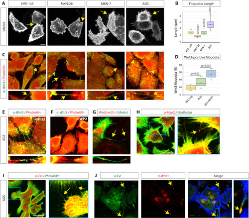
(A) Confocal images of normal gastric epithelial cell line (HFE-145) and gastric cancer (GC) cell lines (MKN-28, MKN-7, and AGS) expressing LifeAct-GFP to visualise actin-based structures. Yellow arrows indicate examples of filopodia. (B) Quantification of filopodia length in GC cell lines MKN-28, MKN-7, and AGS (n=7, 8, 25; n=number of cells). Significance is calculated by Student’s t-test. (C) Immunofluorescent images of HFE, MKN-28, MKN-7, and AGS, stained with antibodies against Wnt3 (green) and actin (Phalloidin-iFluor594, red). Scale bar 10 µm. High-magnification images indicate an example of a Wnt3-bearing cytonemes. Scale bar 2.5 µm. (D) Quantification of Wnt3-positive filopodia in gastric epithelial (HFE-145) and cancer (AGS) cells as a percentage of total filopodia (number of cells analysed = 6, 8, 6). Significance is calculated by Student’s t-test. (E) Immunohistochemistry (IHC) images of AGS cells overexpressing Wnt3 and stained with an antibody against Wnt3 (green) and actin (iFluor594, red). Scale bar 10µm. High-magnification images highlight cytonemes. Scale bar 2.5 µm. (F) IHC images of AGS cells treated with the Porcupine inhibitor IWP2 (100 µM, 48 hr) and stained with an antibody against Wnt3 (green) and actin (iFluor594, red). (G) Live confocal cell imaging of AGS cells expressing Wnt3-mCherry and LifeAct-GFP. Cytoneme-localised Wnt3-mCherry highlighted by yellow arrows. (H–J) IHC images of AGS cells stained with antibodies against (H) Myosin-X (MyoX) and (I) Evi/Wntless (red) and Wnt3 (red) and (J) Evi/Wntless (green Scale bars 10 µm). Phalloidin labels actin (FITC-Phalloidin, green; Phalloidin-iFluor350, blue).
|
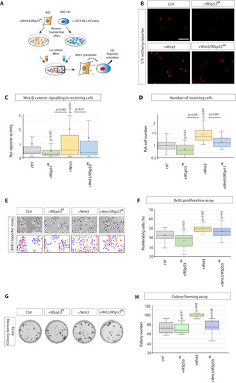
(A) Experimental protocol for measuring paracrine Wnt signalling activation. HFE cells expressing the SuperTOPFlash (STF) reporter, 7×TCF-NLS-mCherry, were cocultivated with AGS cells expressing indicated constructs. Fluorescence of STF mCherry reporter was measured after 48 hr and compared to untransfected control cells. (B) Representative images of STF reporter fluorescence for indicated conditions. Scale bar 100 µm. (C) Quantification of STF mCherry reporter fluorescence in HFE cells co-cultured with AGS (n per condition = 322, 394, 258, and 275). (D) Relative number of HFE cells per image after co-culture with AGS cells expressing indicated constructs. Significance calculated by Student’s t-test with Bonferroni correction for multiple comparisons. (n per condition = 28, 26, 27, and 17; n=number of images). (E) Representative images of proliferating, BrdU-stained (red); co-cultured AGS and HFE-145 cells, as described in (a). Cells were counterstained with haematoxylin (blue dots). Scale bar 100 µm. Complementary images show BrdU+ cells with red dots; blue dots mark BrdU- cells. (F) Quantification of BrdU-stained cells as a percentage of the population. Significance calculated by Student’s t-test with Bonferroni correction for multiple comparisons. (n per condition = 20, 20, 20, and 20; n=number of images). (G), Colony-forming assay of AGS cells. AGS cells were transfected with the indicated constructs and co-cultured with AGS-RFP cells for 2 days. After sorting, AGS-RFP expressing cells were plated at clonal density for 10–12 days; (H), Quantification of spherical colonies. Significance is calculated by Student’s t-test (n=9).
|
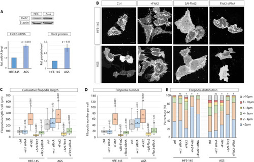
(A) Flot2 protein levels in HFE-145 and AGS cells as quantified by Western blot after normalising to beta-actin levels (n=3) and by RT-qPCR after normalising to Glyceraldehyde-3-Phosphate Dehydrogenase (GAPDH) as a housekeeping gene (n=4). Relative protein and mRNA levels are compared to HFE-145. Error bars represent SEM. Significance is calculated by Student’s t-test. (B) Representative images of HFE and AGS cells expressing membrane-mCherry and indicated Flotillin-2 (Flot2) constructs or siRNA after 48 hr. Scale bars 10 µm. (C–D) Filopodia quantifications of HFE and AGS cells transfected with indicated Flot2 plasmids or siRNA. Significance calculated by Student’s t-test with Bonferroni correction for multiple comparisons. Average cumulative filopodia length (C), average filopodia number per cell (D). (n per condition [HFE]=22, 19, 25, 23, 24). (n per condition [AGS]=25, 21, 25, 25, 25; n=number of cells measured). (E) Distribution of filopodia, categorised by length as a percentage of total filopodia per HFE or AGS cell 48 hr post-transfection with indicated Flot2 plasmids or siRNA. A Pearson’s χ2 test was performed to test for significance between control (ctrl) group (expected) and experimental groups (observed) with 5 degrees of freedom (df) and a p-value <0.05. The specific χ2 values are as follows, HFE: ctrl siRNA 0.86, Flot2 0.001, dnFlot2 <0.001, Flot2 siRNA <0.001, and for AGS: ctrl siRNA 0.65, Flot2 0.007, dnFlot2 <0.001, and Flot2 siRNA <0.001. Asterisks mark significant differences.
|

< (A) Immunohistochemistry (IHC) analysis showing endogenous localisation of Flot2 (green) in AGS cells. TRITC phalloidin was used to visualise actin. Arrows indicate the localisation of Flot2 to filopodia. Scale bars 5 µm. High-magnification images indicate an example of a Flot2-bearing cytonemes. Scale bars 2.5 µm. (B), IHC analysis shows that Flot2 co-localises with Wnt3 on cytonemes. (C) Confocal images showing the subcellular localisation of Flot2-GFP in AGS cells. Arrows indicate the localisation of Flot2-GFP on cytonemes. (D) Confocal images highlighting co-localisation of Flot2-GFP and Wnt3-mCh on cytonemes in AGS cells (arrows). Flot2-GFP and Wnt3-mCherry also cluster and co-localising at a cytoneme contact point (arrow). (E) Representative images of SuperTOPFlash (STF) reporter fluorescence for indicated conditions. Scale bar 100 uM. (F) Relative quantification of STF mCherry reporter fluorescence in HFE cells co-cultivated with AGS cells expressing indicated constructs. Quantifications are relative to AGS control. (n per condition = 322, 443, 403, 258, 336, 306, and 297; n=number of nuclei measured). (G) Relative number of HFE cells per image after co-cultivation with AGS cells expressing indicated constructs. Significance calculated by Student’s t-test with Bonferroni correction for multiple comparisons. (n per condition = 28, 27, 26, 27, 22, 24, and 15; n=number of images). (H) Quantification of BrdU-stained cells as a percentage of the population, after co-cultivation of AGS and HFE cells, as described in Figure 2a. significance calculated by Student’s t-test with Bonferroni correction for multiple comparisons. (n per condition = 20; n=number of images). (I), Colony-forming assay of AGS cells. AGS cells were transfected with the indicated constructs and co-cultivated with AGS-RFP cells for 2 days. After sorting, AGS-RFP expressing cells were plated at clonal density; (J), Quantification of colonies after 10–12 days. Significance is calculated by Student’s t-test (n=9).
|

(A) Immunohistochemistry (IHC) analysis of AGS cells stained for Ror2 (red) and Flot2 (green). Flot2 and Ror2 show co-localisation with a Pearson’s correlation coefficient (PCC) of 0.65 (n=10), highlighted at the membrane by arrows. Scale bars represent 10 µm, and in high-magnification images, 2.5 µm (right). (B–D), Confocal live-cell imaging of AGS cells expressing Ror2-BFP with Flot2-GFP (B), Flot2 siRNA (c) or ∆N-Flot2-GFP (D) and memCherry. Arrows highlight subcellular regions of co-localisations. (E) Live confocal images of AGS cells expressing Ror2-mCherry and indicated organelle markers +/- Flot2 siRNA. Arrows highlight the co-localisation of Ror2-mCherry and mTurq2-Golgi. Scale bar 10 µm F, Quantification of co-localisation of Ror2-mCherry with indicated markers, assessed by PCC. Significance is calculated by Student’s t-test. (n per condition [WT]=7, 10, 8, and 10) (n per condition [Flot2 siRNA]=7, 6, 7, 8). (G) Representative images of AGS cells stably expressing the JNK-KTR-mCherry reporter and indicated constructs after 48 hr. Blue dotted line encircles the cytoplasm and yellow dotted line the nucleus of a representative cell. Scale bar 20 µm. (H) Quantification of the JNK-KTR-mCherry reporter. Nuclear and cytoplasmic fluorescence of cells were measured, and the cytoplasmic: nuclear ratio was calculated. Significance is calculated by one-way ANOVA with Bonferroni correction for multiple comparisons. (n per condition = 136, 109, 74, 109, 82, and 79). (I) Representative confocal images of AGS cells expressing memCherry and indicated constructs for 48 hr. Scale bars 10 µm. (J, K) Quantification of cytoneme length and number of AGS cells transfected with constructs indicated in (i). Significance calculated by Student’s t-test with Bonferroni correction for multiple comparisons. (n per condition = 25, 22, 21, 23, 25, and 21; n=number of cells).
|

(A) Experimental setting to generate small clones expressing indicated constructs in the zebrafish embryo. (B, C) Confocal images of zebrafish epiblast cells injected with Flot2-GFP / memCherry and Flot2-GFP / Wnt8a-mCherry and imaged at 8 hpf. Scale bars represent 10 µm (D) Representative images of zebrafish epiblast cells injected with memCherry indicated constructs. Scale bar 10 µm. (E), (F), Quantification of filopodia from epiblast cells injected. Significance is calculated by Student’s t-test. (n per condition = 17, 20, and 14). (G), Experimental strategy to generate Wnt8a-GFP/mCherry cell clones in F0 Flot1b/2 a Crispants background. (H), Confocal images of zebrafish epiblast cells expressing indicated constructs. (I) Quantification of cytoneme length and number in injected epiblast cells. Significance is calculated by Student’s t-test. (n per condition = 18, 15) (J) In situ hybridisation against pax6a in zebrafish embryos at 30hpf after microinjection of 100 ng/µl of indicated DNA constructs and imaged. Scale bar represents 100 µm. (K) Quantification of forebrain and midbrain primordia length in zebrafish embryos injected as in (A). Significance is calculated by Student’s t-test. (n per condition = 23, 16, 27, 18, and 18; n=number of embryos). (L) Qualitative analysis of phenotype severity in zebrafish embryos injected as indicated in (A). Phenotypes are classified into the categories normal, mild and severe. Numbers in bars represent percentages of total embryos. A Pearson’s χ2 test revealed a significant difference between the ctrl group (expected) and experimental groups (observed) with 2 degrees of freedom (df) and a p-value < 0.05 of χ2 <0.001. Distribution comparison of flot2 injected embryos revealed a significant difference to the wnt8a group (χ2=0.002) and the flot2 + wnt8 a group (χ2<0.001), but not to wnt8a+ΔN-flot2 (χ2=0.28). Asterisks mark significant differences.
|