FIGURE SUMMARY
Title

Vangl2 promotes the formation of long cytonemes to enable distant Wnt/β-catenin signaling

Authors
Brunt, L., Greicius, G., Rogers, S., Evans, B.D., Virshup, D.M., Wedgwood, K.C.A., Scholpp, S.
Source
Full text @ Nat. Commun.

Vangl2 is present on the tips of Wnt8a positive cytoneme.

AF PAC2 zebrafish fibroblasts transfected with indicated constructs and analysed live at 24 h of post-transfection. Yellow arrows indicate Wnt8a on tips of cytonemes. White scale bar equals 5 μm. (n = 25, 9, 14, 6, 25, and 14 cells). G Fluorescent intensity measurements were recorded (HL) along cytoneme length as illustrated in G, from tip at 0–4.5 μm along cytoneme. Cytonemes fluorescent intensity measurements were taken up to 4.5 μm from the cytoneme tip in each case. HL Relative fluorescent intensity analysis (gray value) of tagged proteins Wnt8a-GFP, Wnt8a-mCherry, Mem-mCherry, Ror2-mCherry, and GFP-Vangl2, relative pixel intensity values were measured along cytoneme length starting at the cytoneme tip, (n = 17, 12, 12, 14, 11, and 12 filopodia). Standard error of the mean (SEM) = 1. MO For the fluorescence correlation spectroscopy (FCS) analysis, a focused laser spot was scanned across the membrane for 16s while the intensity was measured as a function of time (G(t)). Auto-correlation curve in red and green for Ror2 and Vangl2, respectively. Cross-correlation curve in gray. Fluorescent cross-correlation spectroscopy (FCCS) revealed cross-correlation of Ror2-mCherry and GFP-Vangl2 when exposed to Wnt5a protein or Wnt8a protein. (n = 5 measurements/condition). CTRL D2 (Ror2) = (37 ± 5) µm2 s−1, D2 (Vangl2) = (23 ± 4) µm2 s−1, KD > 1100 nM; Wnt5a D2 (Ror2) = (14 ± 3) µm2 s−1, D2 (Vangl2) = (19 ± 5) µm2 s−1, KD = 173 nM; Wnt8a: D2 (Ror2) = (11 ± 2) µm2 s−1, D2 (Vangl2) = (16 ± 3) µm2 s−1, KD = 138 nM. Source data are provided as a Source Data file.

Vangl2 controls the emergence of Wnt8a cytonemes in fibroblasts.

AJ Wnt8a-positive PAC2 zebrafish fibroblasts transfected with indicated constructs, imaged and analysed live 24 h of post-transfection. Yellow arrows indicate examples of Wnt8a positive cytonemes. Scale bar = 10 µm. K Number of Wnt8a positive cytonemes per cell (n = number). (n per condition = 25, 9, 14, 6, 25, 14, 31, 36, 13 cells). L Length of Wnt8a positive cytonemes in PAC2 cells (µm). (n per condition = 139, 52, 32, 21, 131, 65, 47, 51, 11 cytonemes). M Breakdown of the percentage of Wnt8a positive cytoneme lengths into 0–5, 5–10, 10–15, 20+ µm categories. Graphs represent mean and standard error of the mean. K, L Two-sided Kruskal–Wallis tests with Bonferroni correction for multiple tests. Statistical significance: p ≤ 0.05. SEM = 1. Corresponding dot plots are shown in Supplementary Fig. 2, and analysis of PAC2 filopodia is shown in Supplementary Figs. 3 and 4. Source data are provided as a Source Data file.

Vangl2 regulates length and contacts of Wnt8a cytonemes in the zebrafish embryo.

A, BTg(vangl2:GFP-Vangl2) zebrafish embryos were injected with mem-mCherry (mCh) or Ror2-Mcherry. A’, B’ 3D renders (3D sr) of A&B at 5hpf. Yellow arrows show examples of Vangl2 positive cytonemes. AAn = 6 embryos. BB’: n = 8 embryos. CG Wild type zebrafish embryos were injected with indicated constructs and imaged at 5hpf. Yellow arrows show examples of Wnt8a positive cytonemes. Blue arrows show examples of thick membrane protrusions. Scale bar = 10 µm. H Number of Wnt8a positive cytonemes per cell (N = number). (n per condition = 3, 6, 3, 9, 7 embryos, n = 87, 123, 44, 154, 93 cells). I Length of Wnt8a positive cytonemes (µm). (n per condition = 81, 269, 54, 82, 24 cytonemes). J Breakdown of the percentage of Wnt8a positive cytoneme lengths into 0–5, 5–10, 10–15, 20+ µm categories. K Wnt8a/Mem-mCh cytoneme with one wnt8a + ve contact point. Scale bar = 10 µm. L Vangl2/Wnt8a/mem-mCh cytoneme with multiple contact points M Quantification of number of Wnt8a-positive contact points per cytoneme. Wnt8a positive points were counted when at tips or at a fixed junction or “turning point” (n = 32, 125, 80, 81, 24 cytonemes). Graphs represent mean and standard error of the mean. Corresponding dot plots are shown in Supplementary Fig. 5. H, I, K Two-sided Kruskal–Wallis tests with Bonferroni correction for multiple tests. Statistical significance: p ≤ 0.05. SEM = 1. Source data are provided as a Source Data file.

Vangl2 activates JNK signaling to control the formation of Wnt8a cytonemes.

A HEK293T cells all transfected with KTR-mCherry and Wnt5a or Wnt8a protein, plus Ror2; Vangl2; Ror2 & Vangl2; Ror2 & ΔN-Vangl2. Wnt8a protein and Wnt5a protein (500 ng/ml) was also added to some conditions 24 h prior to imaging. Blue circle = nuclear, yellow circle = cytoplasmic. B Violin plot of normalized ratio of cytoplasmic to nuclear signal of KTR-mCherry. Increased ratio indicates increasing JNK activity. (n = 69, 32, 32, 25, 30, 24, 43, 75, 60, 49 cells). Two-sided Kruskal–Wallis tests with Bonferroni correction. SEM = 1. C Time series of PAC2 cells at 0 (0‘), 10, 20, 30, 60, and 120 min in control cells and cells post 20 µM SP600125 treatment. D Relative number of filopodia per cell in relation to time = 0 h, at 0, 60, 120 min. (n = 3, 6, 10, 4 cells. n = filopodia at 0, 1, 2 h = (102/111/109, 251/156/122, 158/185/215, 58/50/44). p values over + Sp600125 60′ & 120′ (green bars) significant to 0′. p value over Vangl2 + SP600125 120′ (purple) significant to Vangl2 0′ (yellow). E Relative filopodia length (µm) in relation to time = 0 h, at 0, 60, 120 min. (n = 3, 6, 13, 4 cells. n = filopodia at 0, 1, 2 h = (102/111/109, 251/156/122, 232/274/295, 58/50/44). p value over + Sp600125 60’ (green) significant to 0′. p values over Vangl2 + SP600125 120′ (purple) significant to Vangl2 0′ (yellow). Corresponding dot plot for D, E shown in Supplementary Fig. 6B. Statistical significance: p ≤ 0.05. D, E Two-sided Kruskal–Wallis tests without Bonferroni correction. SEM = 1. Scale bar = 10 µm. Source data are provided as a Source Data file.

Vangl2 activity in the Wnt source cells regulates paracrine Wnt/β-catenin signal activation.

A Schematic of SuperTOP Flash (STF) reporter co-cultivation assay in AGS cells. AGS cells transfected with STF reporter were co-cultivated AGS cells transfected with combinations of with Wnt8a, Vangl2, Ror2 and ΔN-Vangl2 plasmid. B AGS cell STF reporter activation at each condition (i–viii). Scale bar = 10 µm. C Relative STF reporter activation in cells when co-cultured with control; Wnt8a; Vangl2; Wnt8a/Vangl2; Wnt8a/ΔN-Vangl2, Wnt8a/Ror2/Vangl2; Wnt8a/Ror2/ΔN-Vangl2 and Wnt8a/Vangl2/Ror23I. (n = 5, 10, 5, 5, 5, 5, 5, 5 repeats). D AGS cell STF reporter activation after IRSp534k addition: (i–iii); control; Wnt8a/Vangl2; Wnt8a/Vangl2/IRSp534k. Scale bar = 10 µm. E Relative STF reporter activation in cells when co-cultured with control; Wnt8a; Vangl2; Wnt8a/Vangl2; Wnt8a/Vangl2/IRSp534k. (n = 20 biological repeats across two independent experiments). C, E Data represented as box and whisker plots. Whiskers define the minimum and maximum values. Bounds of box indicate the 25th and 75th percentile, center line indicates the median, C calculated with exclusive median. C Student’s t-test and E One-way ANOVA test plus Tukey’s post-hoc test. Statistical significance: p ≤ 0.05. Source data are provided as a Source Data file.

Vangl function in murine telocytes is required for the formation of Wnt deficient intestinal crypt.

A Telocytes stained with FITC-phalloidin. Yellow arrows indicate filopodia protrusions. Blue arrows indicate thicker protrusions. Scale bar = 10 µm. B, C Number of filopodia per cell and average length of filopodia in telocytes treated with control scrambled siRNA and siRNA for Vangl1, Vangl2 and Vangl1/Vangl2, IRSp53. B (n = 35, 21, 22, 27, 6 cells). C (n = 61, 50, 30, 30, 6 cells, n = 3488, 1796, 1964, 1079, 92 filopodia). Graphs represent mean and SEM. Statistical significance: p ≤ 0.05. B, C Two-sided Kruskal-Wallis tests with Bonferroni correction for multiple tests. SEM = 1. D Organoid formation assay: PDGFRα-cre/Rosa2-mTmG telocytes (green), co-cultured with Porcn−/−- intestinal epithelial crypt cells (brightfield) in scrambled siRNA, Vangl1, Vangl2, Vangl1/Vangl2, and IRSP53 siRNA treated conditions. Micrographs were taken day 1 after co-culture, indicating that cells targeted with control or specific siRNAs were establishing extensive contacts with organoid epithelial cells. Arrows indicate long filopodia-like membrane protrusions in siRNA treated control group while in groups treated with Vangl1/2 or IRSp53 siRNA contacts are established via lamellipodia and thicker, telome-like structures. Scale bar = 10 µm. E Organoid formation assay of Porcn deficient crypt cells co-cultured with murine telocytes treated with control scrambled, Vangl1, Vangl2, or Vangl1/Vangl2 siRNA. Organoid counts were recorded per condition (n = 4 assays per condition). F Organoid formation assay of Porcn deficient crypt cells co-cultured with murine telocytes treated with control scrambled, IRSP53 siRNA. (n = 4 assays per condition). Source data are provided as a Source Data file.

Agent-based modeling predicts an essential role for Vangl2 in Wnt-mediated tissue patterning during zebrafish gastrulation.

A, B Comparison of Wnt protein distribution in control zebrafish embryos with zebrafish embryos with Vangl2/Wnt8a overexpression. State of one realization of the model at the indicated time point (t): (Sphere stage = 4 hpf (hours post fertilization); 50% epiboly = 5.3 hpf; 80% epiboly = 8.5 hpf) for base parameter values and Wnt8a and Vangl2 over-expression parameters. Scale bar = 250 µm. For the 80% epiboly panel, the fate of the cells is also plotted in B. Source cells (S), are indicated in red. R = Receiving cells. The colors of the cells in anterior tissue correspond to the relative level of Wnt8 protein received (A). For ease of viewing, Wnt8a protein values have been normalized by the maximum value attained by any cell across the simulation and log transformed. In the cell fate diagram (B), cells acquiring a hindbrain (HB) fate are marked in dark blue, whilst those not acquiring a hindbrain fate are marked in gray. The orange line marks the estimated midbrain-hindbrain boundary (MHB). C Tissue growth properties—a single simulation. Note that the mechanics of the tissue growth are preserved across all conditions and so this graph is representative of all simulations. The red curve shows the proportion of the yolk that is covered by the tissue at the indicated times. The blue curve shows the mean tissue thickness (in terms of the number of cells). D Evolution of the cell number. E, F Histograms of cytoneme lengths at the final state of the simulation (normalized by cell diameter) for the control parameters (E) and with longer cytonemes (F). The gray curve shows a fit of the data to a log-normal distribution with means 2.0 (E) and 3.8 (F). GK Distributions of cell fates over the angular polar coordinate at the end state of the simulation according to a hindbrain Wnt8a threshold of 100 (AU) for control parameters. The length of an arc of a circle is equal to ∅ is equal to 250 µm (G), Wnt8a overexpression parameters (H), longer cytonemes (I), longer Wnt8a positive cytonemes (J), long but fewer Wnt8a positive cytonemes (K). The dark and light blue curves respectively show the mean and standard deviations of the proportion of cell fates acquiring a hindbrain fate in 100 equi-spaced bins around the yolk over 100 model simulations for each condition. The orange line shows the estimated position of the MHB. FB/MB forebrain/midbrain. The red shaded area marks the position of the margin of wnt8a producing cells. Source data are provided as a Source Data file.

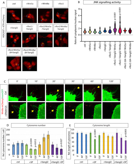
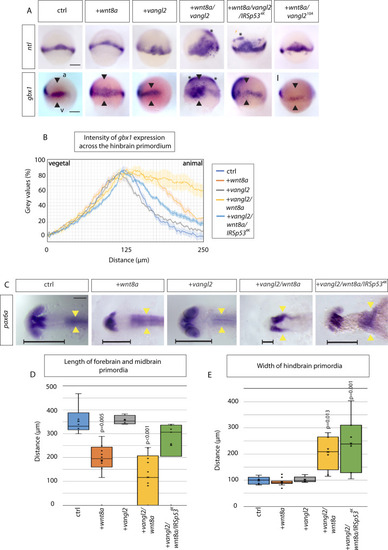
Vangl2 function is crucial for anteroposterior patterning of the zebrafish neural plate.

A In situ hybridization analysis of indicated markers in 60% epiboly zebrafish embryos (6.5 hpf) injected with Wnt8a, Vangl2 and Wnt8a/Vangl2, wnt8a/vangl2/IRSp534K, wnt8a/vangl210A. Scale bar indicates 200μm. (ntl: n = 35, 16, 6, 12, 13, 19 embryos) (gbx1: n = 6, 19, 3, 11, 22, 19 embryos). a = animal pole, v = vegetal pole. Black arrowheads indicate width of gbx1 expression. Asterisks indicate ectopic expression. B Intensity of gbx1 expression from in situ hybridization (gray values in %) across the hindbrain primordium from vegetal (v) to animal (a) pole in embryos injected with indicated constructs at 60% epiboly (6.5 hpf): Control- light blue, wnt8- orange, vangl2 - gray, wnt8a/vangl2- yellow, wnt8a/vangl2/IRSp534K- dark blue. C In situ hybridization to mark pax6a expression in the primordia of the forebrain and the hindbrain in embryos injected with mRNAs for the indicated constructs at 24 hpf: control, wnt8a, vangl2, wnt8a/vangl2, and wnt8a/vangl2/IRSp534K. Scale bar indicates 100 μm. Horizontal black line indicates length of forebrain and midbrain primordium. Yellow arrowheads show the width of the hindbrain primordium, indicating the extent of convergent and extension of these cells. (n = 13, 19, 8, 10, 7 embryos). D Box and whisker plot of length of forebrain and midbrain primordia in control (blue), Wnt8a (orange), Vangl2 (gray), Wnt8a/Vangl2 (yellow) and Wnt8a/Vangl2/IRSp534K (green) 24 hpf larvae (μm). Measured from anterior forebrain pax6a expression to the position of the midbrain-hindbrain boundary (MHB) shown by horizontal black line (C). (n = 6, 12, 4, 10, 7 embryos). E Box and whisker plot of maximum width of hindbrain primordia in control (blue), Wnt8a (orange), Vangl2 (gray), Wnt8a/Vangl2 (yellow) and Wnt8a/Vangl2/IRSp534K (green) 24hpf larvae (μm). Measured from maximum width of hindbrain pax6a expression shown by yellow arrowheads (C). (n = 5, 11, 5, 8, 7 embryos). D, E Data represented as box and whisker plots. Whiskers define the minimum and maximum values. Bounds of box indicate the 25th and 75th percentile. center line indicates the median. Cross indicates mean. Outliers and inner points shown. Statistical significance: p ≤ 0.05. D, E One-way ANOVA tests plus Tukey’s post-hoc test. SEM = 1. Source data are provided as a Source Data file.

Acknowledgments
This image is the copyrighted work of the attributed author or publisher, and ZFIN has permission only to display this image to its users. Additional permissions should be obtained from the applicable author or publisher of the image. Full text @ Nat. Commun.