
- Title
-
Generation of Knockout and Transgenic Zebrafish to Characterize Abcc4 Functions in Detoxification and Efflux of Lead
- Authors
- Lu, X., Long, Y., Li, X., Zhang, L., Li, Q., Wen, H., Zhong, S., Cui, Z.
- Source
- Full text @ Int. J. Mol. Sci.
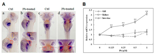
Lead (Pb)-induced expression of the abcc4 gene in developing embryos and adult tissues of zebrafish. (A) Induced expression of the zebrafish abcc4 gene in developing embryos under lead stress. Embryos were treated with 5 μM lead from 24 to 120 h post-fertilization (hpf) and subjected to whole-mount RNA in situ hybridization (WISH) with antisense RNA probe. (a,b) Lateral views; (c,d) dorsal views. Yellow, white and red dashed lines indicate the oral cavity, gill and pronephric tubules, respectively. (B) Transcriptional responses of the zebrafish abcc4 gene to lead treatments in adult tissues. Adult zebrafish were exposed to various concentrations of lead for 24 h. Gills and intestines were taken from four females, and kidneys were taken from four females and four males. The same tissues from different fish were mixed for isolation of total RNA and subjected to qRT-PCR analysis of abcc4 mRNA levels relative to the control. One-way analysis of variance (ANOVA) followed by Duncan’s post-hoc test was performed, and the symbol above the error bars indicates a significant difference (* p < 0.05 and ** p < 0.01) of mRNA levels in the same tissue among the indicated doses of treatments. |
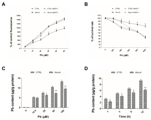
Cellular functions of zebrafish Abcc4 in lead excretion. (A) The contents of monochlorobimane (MCB) in LLC-PK1 cells expressing zebrafish Abcc4 and transfected empty vectors (CTRL) with or without 25 μM MK571 were determined with dye accumulation assays after exposure to lead at the indicated concentrations for 24 h. (B) Survival rates of LLC-PK1 cells expressing zebrafish Abcc4 and CTRL with or without 25 μM MK571 were determined with 3-[4, 5-dimethylthiazol-2-yl]-2, 5 diphenyl tetrazolium bromide (MTT) assays after exposure to lead at the indicated concentrations for 24 h. (C) Contents of lead in LLC-PK1 cells expressing Abcc4 and CTRL after exposure to lead at the indicated concentrations for 12 h. (D) Contents of lead in LLC-PK1 cells expressing Abcc4 and CTRL after exposure to 50 μM lead at the indicated time points. Values are expressed as means ± SD (n = 3). Significant differences are indicated as * p < 0.05 and ** p < 0.01. |

Generation of zebrafish abcc4−/− mutants with a clustered regularly interspaced palindromic repeats (CRISPR)/Cas9 system. The red color indicates the sgRNA target sequences for zebrafish abcc4 gene. (A) Diagram of the target site in the zebrafish abcc4 genome. (B) Abcc4 amino acids of wild-type (WT) and abcc4−/− mutants. (C) The predicted truncation of zebrafish Abcc4 protein. N and C indicate N terminal and C terminal, respectively. The red star symbol indicates a sgRNA targeting site of the abcc4 gene. |

Identification and functional characterization of zebrafish abcc4−/− mutants. (A) PCR amplification of WT and mutant F3 zebrafish. M: Molecular markers; WT: wild type zebrafish; F3+/−: heterozygous mutants. Red arrows point to the specific DNA bands for mutants. (B) Sequencing maps of WT and homozygous mutants. Oval frame: the 5-bp (CATGC) deletion in homozygotes. (C) The expression of Abcc4 protein in WT, abcc4+/− and abcc4−/− larvae at 96 hpf detected with Western blotting. (D) Survival rates of abcc4−/− and WT larvae were monitored after exposure to 400 μM lead from 96 to 168 hpf. (E) Lead contents in abcc4−/− and WT larvae after treatment with 10 μM lead at the indicated exposure time points. Values are expressed as means ± SD (n = 3). Significant differences are indicated by * p < 0.05 and ** p < 0.01. PHENOTYPE:
|

Expression of foreign Abcc4 in transgenic zebrafish. (A) A primer pair (Flag-F and ABC-R3) was designed to recognize the abcc4 coding sequence (CDS) and Flag sequence. (B) Positive F2 fish were individually crossed with WT fish to obtain positive F3 embryos. Sixty larvae at 96 hpf were pooled for RNA extraction and subsequent RT-PCR analysis with a primer pair (Flag-F and ABC-R3). WT: Wild-type zebrafish embryos at the same developmental stage. F3 abcc4-1# and F3 abcc4-2# indicated the pooled larval zebrafish at 96 hpf from two positive F2 zebrafish, which were individually crossed with WT fish, respectively. (C) qPCR analysis for abcc4 mRNA levels with a primer pair (abcc4–qPCR-F and abcc4–qPCR-R). (D) Western blotting analysis of Flag-tagged Abcc4 expression in F3 embryos. (E) WISH analysis of abcc4 expression in WT and transgenic zebrafish larvae. Arrow heads indicate the brain and intestine. |

Functional characterization of Abcc4 in lead detoxification in transgenic zebrafish. (A) Survival rates of abcc4-transgenic and WT embryos were monitored after treatment with different concentrations of lead (0–600 μM) from 96 to 168 hpf. (B) Survival rates of abcc4-transgenic and WT larvae were detected after exposure to 400 μM lead for 72 h. (C) Contents of lead in abcc4-transgenic and WT larvae after exposure to 10 μM lead with or without 1 mM glutathione (GSH) at the indicated exposure time points. (D) Contents of lead in abcc4-transgenic and WT larvae at the indicated exposure time points after treatment with a medium containing 10 μM lead and simultaneously with 5 μM buthionine sulfoximine (BSO), an inhibitor of GSH biosynthesis. (E) Intracellular GSH contents in abcc4-transgenic and WT larvae exposed to 10 μM lead at the indicated exposure time points. (F) ATPase activities as shown by inorganic phosphate (Pi) levels in abcc4-transgenic and WT larvae after treatment with 10 μM lead at the indicated exposure time points. Values are expressed as means ± SD (n = 3). Significant differences are indicated by * p < 0.05 and ** p < 0.01. PHENOTYPE:
|