
- Title
-
Atrial and Sinoatrial Node Development in the Zebrafish Heart
- Authors
- Martin, K.E., Waxman, J.S.
- Source
- Full text @ J Cardiovasc Dev Dis
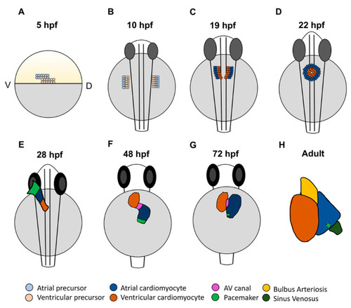
Stages of zebrafish heart development. (A) At 5 h post-fertilization (hpf), cardiac progenitors are located in the lateral marginal zone, with atrial progenitors located more ventrally than ventricular progenitors. (B) Following gastrulation at the tailbud stage (10 hpf), cardiac progenitors migrate to the anterior lateral plate mesoderm (ALPM). (C) In the ALPM, progenitors begin to differentiate and express chamber-specific genes. (D) Cells then migrate to the midline and fuse, forming the cardiac disc where atrial cardiomyocytes surround ventricular cardiomyocytes. (E) The disc elongates to form the linear heart tube, which begins beating by 24 hpf. At 28 hpf, the dominant pacemaker covers a large area at the venous pole. (F) By 48 hpf, the heart has finished looping and the two chambers have formed. Here, the pacemaker is a ring at the venous pole. (G) By 72 hpf, the dominant pacemaker is restricted to a small population of cells in the inner curvature at the venous pole of the atrium. (H) In the adult heart, the bulbus arteriosus and sinus venosus, which serve as the outflow and inflow tracts, respectively, have matured. The dominant pacemaker is located at the sinus venosus–atrial junction. |

Signaling pathways required for different stages of atrial development and cardiac maintenance in zebrafish. (A) Pathways required for specification of chamber progenitors in the early embryo. (B) Factors responsible for differentiation of atrial cardiomyocytes within the ALPM and at the venous pole. (C) Genes shown to promote or repress atrial identity within differentiated embryonic ventricular cardiomyocytes and repress ventricular gene expression in atrial cardiomyocytes. A—atrium, V—ventricle. |
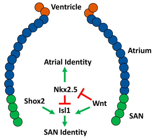
Molecular network shown to be responsible for differentiation of the sinoatrial node (SAN) in embryonic zebrafish. |