FIGURE SUMMARY
Title

Unbiased Identification of Angiogenin as an Endogenous Antimicrobial Protein With Activity Against Virulent Mycobacterium tuberculosis

Authors
Noschka, R., Gerbl, F., Löffler, F., Kubis, J., Rodríguez, A.A., Mayer, D., Grieshober, M., Holch, A., Raasholm, M., Forssmann, W.G., Spellerberg, B., Wiese, S., Weidinger, G., Ständker, L., Stenger, S.
Source
Full text @ Front Microbiol

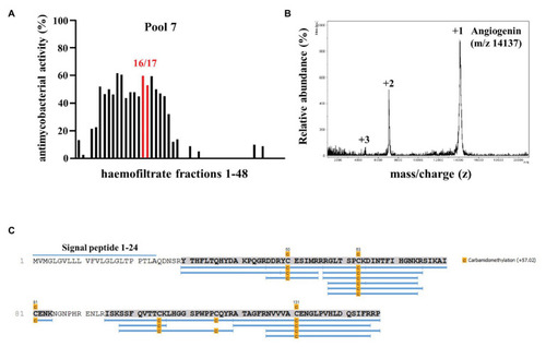
Screening a hemofiltrate library for activity against extracellular Mycobacterium tuberculosis (Mtb). (A) Around 48 fractions (each 10 μl) of pool seven of the hemofiltrate library were incubated for 96 h with extracellular Mtb. 3H-Uracil was added for the final 24 h of incubation. The uptake of 3H-Uracil was measured by scintillation counting in a ß-counter. Antimicrobial activity was calculated by comparison to the untreated control. The bars present the average antimycobacterial activity (%) calculated from triplicates of a single experiment. (B) Subfractions 7–16/17 were analyzed via mass spectrometry. Spectrum analysis yielded three distinct peaks, identifying Angiogenin with a molecular mass of 14,137 Dalton. (C) Angiogenin sequence coverage (88%) by MS/MS analysis of the sample after carbamidomethylation and digestion with trypsin. Full sequence (Angiogenin precursor): signal peptide: 1–24 and Angiogenin 25–147.

Activity of synthetic Angiogenin against extracellular Mtb. About 2 × 10⁶ extracellular Mtb were incubated for 96 h with synthetic Angiogenin (PSL Heidelberg). 3H-Uracil was added for the final 24 h, of incubation and 3H-Uracil uptake was measured by scintillation counting. Antimycobacterial activity was calculated as follows: [(counts/sample: count/untreated control) ×100]. Bars represent the mean antibacterial activity (%) ± SD calculated from triplicates of three independent experiments.

Effect of Angiogenin on macrophage viability and Mtb multiplication. (A) Macrophages were incubated for 24 h with Angiogenin (10 μM, PSL Heidelberg) or diluent. Macrophage viability was quantified using a PrestoBlue™ assay and is given in % of the un-treated sample. Bars represent the mean viability of macrophages (%) ± SD calculated from triplicates of three independent experiments. (B) Macrophages were infected with Mtb, followed by incubation with Angiogenin (10 μM) for 4 days. Lysates were plated on 7H11 agar plates and the number of CFU counted after 21 days of incubation. Mtb-multiplication was calculated by comparison of the CFU determined at d4 and d0. The graph gives the mean values ± SD of n-fold mycobacterial growth as compared to d0 for six independent donors. Statistical analysis was performed using a paired t-test.

In silico design of Angie1 from Angiogenin. (A) Sequence of Angiogenin precursor (1–24: signal peptide; 25–147: Angiogenin). The cytolytic active site was determined by AMPA (64–80aa) and is highlighted in green. (B) Helical wheel projection of the cytolytic region (left panel) demonstrating the polarity of the amino acids. The right projection shows the modifications of the cytolytic region yielding Angie1. (C) Mass spectrometry analysis of Angie1. Angie1 was analyzed by LC-ESI-MSMS (Orbitrap Elite system). The spectrum shows the [M+2H]2+, [M+3H]3+, and [M+4H]4+ multicharged signals with the monoisotopic m/z value.

Antimycobacterial activity of Angie1 against extracellular Mtb. About 2 × 10⁶ extracellular Mtb were incubated for 96 h with Angie1 (CFP). Uptake of 3H-Uracil was measured by scintillation counting. Antimicrobial activity was calculated by comparison of treated samples to untreated controls. Bars represent the mean antibacterial activity (%) ± SD calculated from triplicates of three independent experiments.

Effect of Angie1 on macrophage viability and multiplication of intracellular Mtb. (A) Macrophages were incubated with Angie1 (PSL Heidelberg) at indicated concentrations for 24 h and viability was determined by the PrestoBlue™ assay. Bars represent the mean viability of macrophages (%) ± SD calculated from triplicates of three independent experiments. (B) Macrophages were infected with Mtb followed by incubation with Angie1 for 4 days. Mtb-multiplication was determined by plating cell lysates on 7H11 agar plates to determine and counting the number of CFU after 21 days of incubation. The graph gives the mean values ± SD of n-fold mycobacterial growth as compared to d0 for four independent donors.

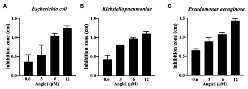
Antimicrobial effect of Angie1 against fast-growing bacteria. About 2 × 10⁷ bacteria were seeded into a petri dish containing agarose. Angie1 (Core Facility Peptidomics) was given into cavities in the agarose. After 3 h, plates were overlaid with agarose and after 18 h of incubation, inhibition zones were measured for (A)Escherichia coli, (B)Klebsiella pneumoniae and (C)Pseudomonas aeruginosa. Bars represent the mean values of inhibition zone size (cm) ± SD calculated from three independent experiments.

Half-life of Angie1 in human serum. Human serum was spiked with Angie1 (10 μM) and aliquots of the samples were harvested at indicated time points. The amount of peptide was determined by mass spectrometry. The graph shows the amount of peptide (%) of the initial inoculum ± SD of three individual measurements.

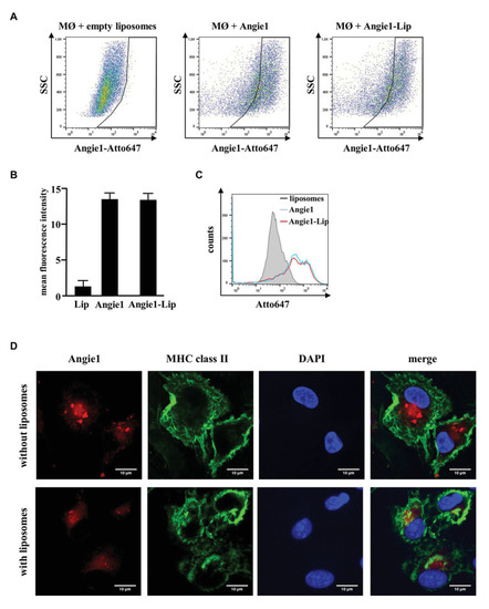
Uptake of Angie1 and Angie1-lip in macrophages. (A) Macrophages were incubated overnight with Atto647N-labeled Angie1 or Angie1-lip (both 54 μM, PSL Heidelberg). After 18 h, cells were harvested and analyzed by flow cytometry. Dot plots show one representative donor of three for each group. (B) The graph shows the mean fluorescence intensity (FI) ± SD of Atto647-positive cells for empty liposomes, Angie1 and Angie1-Lip of three independent donors. Statistical analysis was performed using a non-parametric Wilcoxon-Rank Test for paired samples. (C) The histogram shows the counts of Atto647-positive populations for empty liposomes, Angie1 and Angie1-Lip for one representative donor of three donors. (D) Macrophages were incubated with Angie1-Atto647N (upper panels) or Angie1-Atto647N-Lip (lower panels). After 18 h, cells were stained for MHC class II. Cell nuclei were stained with DAPI. Images were acquired using an inverted laser scanning confocal microscope (Zeiss LSM 710). Depicted images show representative area of one out of three donors with similar result.

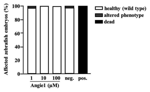
Angie1 is not toxic to zebrafish embryos. Zebrafish embryos were scored for mortality or altered phenotypes at 48 h post fertilization (hpf) after exposure for 24 h to Angie1 at the indicated concentrations or the negative control (PBS) or positive control [NRC-03 antimicrobial peptide, (AMP)]. Altered phenotypes include necrosis and non-lethal lysis (cytotoxicity), heart edema, reduced or absent circulation (cardiotoxicity), delayed development or malformations (developmental toxicity), and reduced or absent touch escape response (neurotoxicity). Note that Angie1 caused no significant toxicity. n = 60 embryos each group.

Acknowledgments
This image is the copyrighted work of the attributed author or publisher, and ZFIN has permission only to display this image to its users. Additional permissions should be obtained from the applicable author or publisher of the image. Full text @ Front Microbiol