Direct targets of BMP signaling in DV axial patterning.

(A) Animal view, dorsal to right of mean pSmad5 intensities at mid-gastrula stage (8 hpf) WT (n = 4) and bmp7 mutant (n = 3) embryos. (B) Differential gene expression in WT and bmp7 mutants at 8 hpf using RNA-seq. Significantly up-regulated genes in WT compared to bmp7 mutants are shown in red, and significantly down-regulated genes are shown in blue. All other genes are shown in gray. A subset of known BMP-dependent genes is highlighted. See Table A in S1 Data for underlying data. (C) Schematic of method to isolate RNA for sequencing from CHX-treated bmp7 mutant embryos injected with BMP2/7 protein. (D) Representative image of pSmad5 intensities of all nuclei of an individual bmp7 mutant (n = 9) and bmp7 mutant injected with 10 pg of BMP2/7 protein (n = 9). Animal pole facing up. (E) Differential gene expression using RNA-seq of CHX-treated bmp7 mutants versus CHX-treated bmp7 mutants injected with BMP2/7 protein. Significantly up-regulated genes with BMP2/7 protein injection are shown in red, and significantly down-regulated genes are shown in blue. All other genes are shown in gray. A subset of BMP direct target genes is highlighted. See Table B in S1 Data for underlying data. (F) GO term analysis for biological processes of the 57 direct target genes. See Table C in S1 Data for underlying data. A.U. is arbitrary units. BMP, Bone Morphogenetic Protein; CHX, cycloheximide; DV, dorsal–ventral; GO, Gene Ontology; hpf, hours post fertilization; pSmad5, phosphorylated Smad5; RNA-seq, RNA sequencing; TGFβ, Transforming Growth Factor Beta; WT, wild-type.

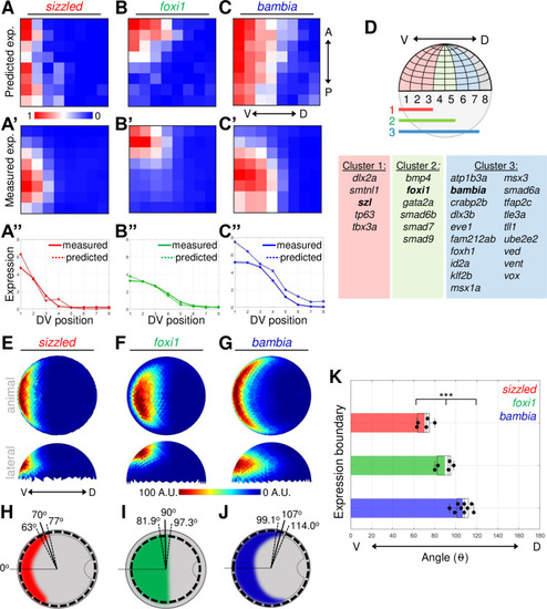
Three distinct expression domains of BMP direct target genes.

(A–C) Heat map of gene expression patterns from Seurat analysis of mid-gastrula (8 hpf) scRNA-seq dataset. Predicted expression normalized across all bins of sizzled(A), foxi1(B), and bambia(C). (A’–C’) Average FISH intensity measured in early gastrula (7 hpf) WT embryos divided into 64 equally spaced bins. (A”–C”) Measured and Seurat predicted expression profiles across the DV axis. Each point is the sum of the expression intensity from all bins at 1 DV position. See Tables A–C in S2 Data for underlying data. (D) Schematic of embryonic grid divided into 64 bins and the 3 nested expression domains. Table of 27 ventrally expressed target genes divided into 3 clusters based on their Seurat expression profiles. (E–G) Animal and lateral views of average FISH signal in WT embryos at early gastrula (7 hpf) of sizzled (n = 5) (E), foxi1 (n = 5) (F), and bambia (n = 9) (G). (H–J) Schematic of animal view (dorsal to right) of expression domains in early gastrula embryos of sizzled(H), foxi1(I), and bambia(J). The mean (solid line) and standard deviation (dotted lines) of expression boundaries shown as degrees across the DV axis. Position of domain boundary measured from the average intensity from a 40-μm band of cells across the DV axis at the location indicated by the dotted circle. (K) DV position of the expression boundaries of individual WT embryos. See Table K in S2 Data for underlying data. ***P < 0.001 in comparing 3 expression domains using 1-way ANOVA. A.U. is arbitrary units. ANOVA, analysis of variance; BMP, Bone Morphogenetic Protein; DV, dorsal–ventral; FISH, fluorescent in situ hybridization; hpf, hours post fertilization; scRNA-seq, single-cell RNA sequencing; WT, wild-type.

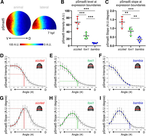
(A) Animal and lateral view of average pSmad5 intensities in early gastrula (7 hpf) WT embryos (n = 5). (B) Measurement of pSmad5 intensity at the expression boundaries of sizzled (red), foxi1 (green), and bambia (blue) across the DV axis of WT embryos at 7 hpf. See Table A in S3 Data for underlying data. (C) Measurement of pSmad5 gradient slope at the expression boundaries of sizzled (red), foxi1 (green), and bambia (blue) across the DV axis of WT embryos at 7 hpf. See Table B in S3 Data for underlying data. (D–F) WT pSmad5 profiles across the DV axis. The intensity is averaged from a 40-μm band of cells around the DV axis at the location shown in red in the right corner embryo schematic of each panel. One WT clutch was used for (D, E, G, H) (n = 5), another WT clutch was used for (F, I) (n = 6). Positions of expression boundaries for sizzled (D), foxi1 (E), and bambia (F) are shown as vertical solid lines. Level of pSmad5 at the boundary is indicated as a horizontal dotted line. Colored dots indicate positions where target genes are expressed. Standard deviations of expression boundaries are shaded. See Tables C–E in S3 Data for underlying data. (G–I) Slopes of pSmad5 profiles are shown in (D–F). Positions of expression boundaries for sizzled (G), foxi1 (H), and bambia (I) are shown as vertical solid lines. Slope of pSmad5 at the boundary is indicated as a horizontal dotted line. Colored dots indicate positions where target genes are expressed. Standard deviations of expression boundaries are shaded. See Tables F–H in S3 Data for underlying data. A.U. is arbitrary units. **P < 0.01, ***P < 0.0001 in comparing pSmad5 levels and slopes using unpaired 2-tailed Student t tests. DV, dorsal–ventral; hpf, hours post fertilization; pSmad5, phosphorylated Smad5; WT, wild-type.

(A) Model of 3 target gene expression domains in WT gastrula embryos positioned by either distinct pSmad5 levels or distinct pSmad5 gradient slopes. (B) Model of predicted target gene expression boundaries in chordin mutant gastrula embryos corresponding to distinct pSmad5 levels or gradient slopes if cells are interpreting pSmad5 concentration or shape, respectively. (C) Animal view of average pSmad5 intensities of early gastrula (7 hpf) WT (n = 6) and chordin mutants (n = 5). (D–F) Animal view of average FISH intensities of WT and chordin mutants for sizzled (D) (WT n = 8, chd-/- n = 8); foxi1 (E) (WT n = 6, chd-/- n = 7); and bambia (F) (WT n = 9, chd-/- n = 4). (G–I) pSmad5 profiles of WT (black solid line) and chordin mutants (black dotted line). Location of 40-μm band of cells that was averaged is indicated on embryo in top right corner. Expression boundaries of sizzled (G), foxi1 (H), and bambia (I) in WT (solid colored line) and chordin mutants (dotted colored line). Standard deviations of expression boundaries are shaded. See Tables A–C in S4 Data for underlying data. (J–L) Slope of pSmad5 profiles shown in (G–I) of WT (black solid line) and chordin mutants (black dotted line). Expression boundaries of sizzled (J), foxi1 (K), and bambia (L) in WT (solid colored line) and chordin mutants (dotted colored line). Standard deviations of expression boundaries are shaded. See Tables D–F in S4 Data for underlying data. A.U. is arbitrary units. FISH, fluorescent in situ hybridization; hpf, hours post fertilization; pSmad5, phosphorylated Smad5; WT, wild-type.

Thirty-minute duration of BMP2/7 sufficient for <italic>sizzled</italic>, <italic>foxi1</italic>, and <italic xmlns:xlink='http://www.w3.org/1999/xlink'>bambia</italic> target gene expression.

(A) Model of target gene expression regulated by distinct pSmad5 levels or distinct durations of BMP signaling. (B) Model of target gene activation after a 30-minute pulse of BMP ligand exposure. If target genes are activated by different pSmad5 levels, then all 3 target genes will be expressed following exposure to high levels of BMP signaling. If differences in signal duration activate BMP target gene expression, then a gene that requires a short signal duration will be expressed, but genes requiring longer signal durations will not be expressed. (C) Experimental schematic of a bmp7 mutant embryo injected with 5 pg of BMP2/7 protein that is fixed 30 minutes postinjection for pSmad5 immunostaining or FISH. (D) Representative immunostaining of pSmad5 intensities of an uninjected bmp7 mutant (n = 8) and a bmp7 mutant injected with 5 pg of BMP2/7 protein (n = 9). Animal pole is facing up. (E–G) Representative FISH in bmp7 mutants uninjected or injected with 5 pg of BMP2/7 protein for sizzled(E) (n = 11 uninjected, n = 11 injected), foxi1(F) (n = 10, n = 11), and bambia(G) (n = 10, n = 11). Animal pole is facing up. A.U. is arbitrary units. BMP, Bone Morphogenetic Protein; FISH, fluorescent in situ hybridization; hpf, hours post fertilization; pSmad5, phosphorylated Smad5; WT, wild-type.

Target genes <italic>sizzled</italic> and <italic>bambia</italic> respond to BMP in a concentration-dependent manner.

(A) Representative pSmad5 immunostaining intensities of disassociated cells from bmp7 mutants and bmp7 mutants treated with 5 or 20 ng/ml BMP2/7 protein. (B, C) Representative FISH for sizzled(B) and bambia(C) in bmp7 mutants and bmp7 mutants treated with 5 or 20 ng/ml BMP2/7 protein. (D) Table displaying the proportion of cells above the predicted pSmad5 threshold for sizzled (60 A.U.) in each condition, and the proportion of cells expressing sizzled in each condition. Cells with greater than 10% of maximum signal intensity are considered to be expressing sizzled. (E) Table displaying the proportion of cells above the predicted pSmad5 threshold for bambia (7 A.U.) in each condition, and the proportion of cells expressing bambia in each condition. Cells with greater than 10% of maximum signal intensity are considered to be expressing bambia. A.U. is arbitrary units. BMP, Bone Morphogenetic Protein; FISH, fluorescent in situ hybridization; pSmad5, phosphorylated Smad5.

Acknowledgments
This image is the copyrighted work of the attributed author or publisher, and ZFIN has permission only to display this image to its users. Additional permissions should be obtained from the applicable author or publisher of the image. Full text @ PLoS Biol.