FIGURE SUMMARY
Title

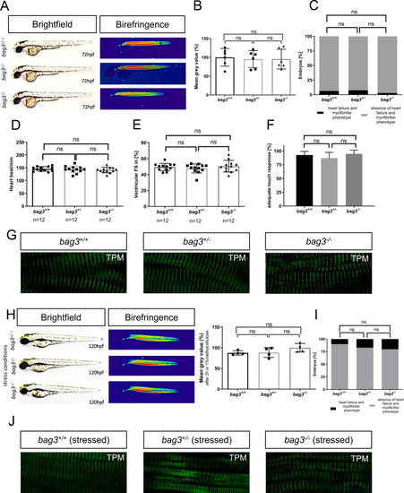
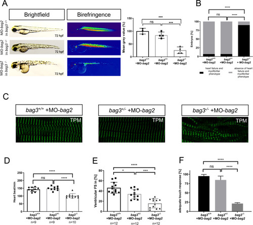
Genetic compensation prevents myopathy and heart failure in an in vivo model of Bag3 deficiency

Authors
Diofano, F., Weinmann, K., Schneider, I., Thiessen, K.D., Rottbauer, W., Just, S.
Source
Full text @ PLoS Genet.

Generation of zebrafish <italic>bag3</italic> knockout by CRISPR/Cas9 gene editing.

(A) Structure of the zebrafish bag3 gene and protein. Exon 2 is the target for the CRISPR/Cas9 gene editing in zebrafish bag3. The CRISPR/Cas9-induced mutation (19 bp deletion) in bag3 is shown in bag3 mutant DNA sequencing chromatogram. The 19 nucleotides deletion in bag3-/- leads to a frame shift, the introduction of a premature stop codon and thereby the premature termination of Bag3 translation, as demonstrated by the alignment of the Bag3-/- and Bag3+/+ aminoacid sequences (only partial aminoacid sequence shown) (B-C) Immunoblot analysis of 72 hpf bag3+/+ embryo protein lysates compared to lysates obtained from bag3-/- clutchmates with antibody against zebrafish Bag3. The figure shows one representative immunoblot from three independent experiments (N = 3, mean ± SD, P<0.0001 determined using two-tailed t-test). (D) Quantitative real-time PCR of bag3+/+ and bag3-/- embryos at 72 hpf shows significant downregulation of bag3 mRNA levels in bag3-/- embryos (N = 3, mean ± SD, P = 0.0004 determined using two-tailed t-test).

EXPRESSION / LABELING:
Gene:
Fish:
Anatomical Term:
Stage: Protruding-mouth
PHENOTYPE:
Fish:
Observed In:
Stage: Protruding-mouth

Genetic loss of <italic>bag3</italic> does not interfere with heart and skeletal muscle function.

(A) Lateral view of brightfield and birefringence images for bag3+/+, bag3+/- and bag3-/- embryos at 72 hpf exhibiting homogenous birefringence signals. (B) Densitometric analysis of birefringence does not report muscle disorganization in bag3-/- embryos. Representative samples are shown (n = 6, One-way ANOVA followed by tukey's multiple comparison analysis, P = 0.9688). (C) Embryos at 72 hpf reveal no myopathic phenotype (N = 3, n = 100, two-tailed value for Fisher´s exact test. bag3+/+ and bag3+/- P = 0.8096; bag3+/+ and bag3-/- P = 0.4949; bag3+/- and bag3-/- P = 0.1925). (D) Heart rate quantification at 72 hpf reveals no impairments under physiological conditions (N = 3, n = 12, mean ± S.D. One-way ANOVA followed by tukey's multiple comparison analysis, P = 0.6026). (E) Quantification of ventricular FS at 72 hpf does not reveal contractile dysfunction (N = 3, n = 12, mean ± S.D. One-way ANOVA followed by tukey's multiple comparison analysis, P = 0.5061). (F) Quantification of the touch evoked assay. bag3-/- embryos reveal no significant difference in responsiveness upon mechanical stimulus (N = 3, n = 90, mean ± S.D. One-way ANOVA followed by tukey's multiple comparison analysis, P = 0.7851). (G) Immunostaining of bag3+/+, bag3+/- and bag3-/- embryos at 72 hpf, with sarcomeric Tropomyosin, showing no muscle fiber disruptions. (H) Brightfield and birefringence images of mutant embryos at 120 hpf under stressed conditions reveal no myopathic phenotype, which was confirmed by densitometric analysis of birefringence signals. Representative samples are shown (n = 4, One-way ANOVA followed by tukey's multiple comparison analysis, P = 0.1824). (I) Embryos, under stressed conditions, do not develop myopathic phenotype (N = 4, n = 50, two-tailed value for Fisher´s exact test. bag3+/+ and bag3+/- P = 0.6050; bag3+/+ and bag3-/- P = 0.8320; bag3+/- and bag3-/- P = 0.8582). (J) Immunostaining of bag3+/+, bag3+/- and bag3-/- embryos at 120 hpf under stressed conditions (2 hours in 1% methylcellulose), with sarcomeric Tropomyosin, showing no muscle fiber disruptions.

Targeted knock-down of Bag3 leads to (cardio-)myopathy in zebrafish (A) Brightfield and birefringence images of MO-bag3 and MO-bag3 5bp mm injected embryos developed (cardio-)myopathy phenotype at 72 hpf. The densitometric analysis of birefringence supports the presence of myopathy phenotype only in bag3 morphants. Representative samples are shown (n = 4, P = 0.0003 determined using two tailed t-test). (B)bag3 splice MO injected embryos develop (cardio-)myopathy phenotype (78.42±9.88%) whereas control-injected embryos developed no pathological phenotype (78.42±9.89%) (N = 3, n = 30/50 mean± SD P<0.0001 determined using two-tailed t-tests). (C) Tropomyosin immunostaining of MO-bag3 and MO-bag3 5bp mm embryos at 72 hpf shows that embryos injected with Bag3 splice MO develop muscle fiber disruptions. (D) Heart rate quantification of bag3 morphants reveals impairments at 72 hpf (N = 3, n = 9/12, P = 0.6026. HR 5bp-mismatch-MO injected embryos: 153±22.98 heart beat/min; HR bag3 morphants: 106±23.58 heart beat/min; mean ± S.D. P<0.0001 determined using two-tailed t-tests). (E) FS of bag3 morphant ventricles at 72 hpf is significantly reduced (16,88±8.56%), compared to MO-bag3 5bp mm injected embryos (FS: 50.48±9.90%) (N = 3, n = 12; Mean± SD P<0.0001 determined using two-tailed t-tests). (F) Touch evoked assay reveals significant reduction in responsiveness upon mechanical stimulus for bag3 morphants (20.78±4.46%) and not for 5bp-mismatch-MO injected embryos (92.78±4.40%) (N = 3, n = 40, mean ± S.D. P = 0.0005, determined using two-tailed t-tests).

Knockdown of Bag3 leads to heart and skeletal muscle dysfunctions only in bag+/+and bag3+/-embryos. (A) Brightfield and birefringence images of bag3+/+, bag3+/- and bag3-/- embryos at 72 hpf injected with MO-bag3. Densitometric analysis of birefringence (n = 4). Representative samples are shown (One-way ANOVA followed by tukey's multiple comparison analysis, P = 0.0012). (B) Only bag3-/- embryos injected with MO-bag3 do not show (cardio-)myopathy, whereas bag3+/+and bag3+/- develop the characteristic bag3 morphant phenotype (N = 3, n = 40, mean ± S.D., P<0.0001, two-tailed value for Fisher´s exact test. bag3+/+ and bag3+/- P = 0.5346; bag3+/+ and bag3-/- P<0.0001; bag3+/- and bag3-/- P<0.0001). (C) Tropomyosin immunostainings of bag3+/+, bag3+/-, and bag3-/- embryos injected with MO-bag3 at 72 hpf reveal that only bag3-/- mutant embryos injected with MO-bag3 does not develop muscle fiber disruptions. (D) Heart rate quantification at 72 hpf reveals impairments only in bag3+/+and bag3+/- embryos injected with MO-bag3 (N = 3, n = 9, mean ± S.D. One-way ANOVA followed by tukey's multiple comparison analysis, P = 0.0008). (E) Quantification of ventricular FS at 72 hpf reveals contractile dysfunctions only in bag3+/+and bag3+/- embryos injected with MO-bag3 (N = 3, n = 9, mean ± S.D. One-way ANOVA followed by tukey's multiple comparison analysis, P<0.0001). (F)bag3-/- embryos injected with MO-bag3 show proper flight response upon mechanical stimulus, whereas bag3+/+and bag3+/- embryos injected with MO-bag3 do not show an adequate touch response (N = 3, n = 60, mean ± S.D. One-way ANOVA followed by tukey's multiple comparison analysis, P<0.0001).

Bag2 mediates genetic compensation in <italic>bag3</italic> knockout zebrafish.

(A) Schematic description of proteomic workflow. Acquired spectra were analyzed against the Uniprot zebrafish database using MaxQuant. (B) Detection levels of label free quantification (LFQ) intensity ratio between bag3-/- and bag3+/+ zebrafish samples of three orthologs of the Bag family protein, Bag1, Bag2 and Bag3. The values are shown as Log2 fold change ≥2 and ≤2. (C) Quantitative real-time PCR of bag3-/- and bag3+/+ zebrafish embryos at 72 hpf shows significant upregulation of bag2 mRNA levels, whereas bag1 transcript levels are not increased (N = 3, mean±S.D, One-way ANOVA followed by tukey's multiple comparison analysis **bag1 vs bag2 P = 0.0057, ** bag2 vs bag3 P = 0.0026). (D) Quantitative real-time PCR of MO-bag3- or MO-bag3 5bp mm-injected embryos shows no upregulation of bag2 mRNA levels in bag3 morphants at 72 hpf. (n = 3, mean±S.D, P = 0.1080 determined using two-tailed t-test).

EXPRESSION / LABELING:
Genes:
Fish:
Knockdown Reagent:
Anatomical Term:
Stage: Protruding-mouth
PHENOTYPE:
Fish:
Observed In:
Stage: Protruding-mouth

Targeted knockdown of <italic>bag2</italic> does not lead to heart and muscles dysfunction in zebrafish embryos.

(A) Brightfield and birefringence images and the densitometric analysis of birefringence (n = 4) of MO-bag2 and MO-bag2 5bp mm injected embryos reveal that bag2 morphants do not develop a (cardio)-myopathy phenotype by 72 hpf. Representative samples are shown (P = 0.7891 determined using two tailed t-test). (B) Tropomyosin immunostainings of MO-bag2- and MO-bag2 5bp mm-injected embryos at 72 hpf demonstrate that bag2 morphants do not develop fiber disruptions. (C)bag2 morphants do not develop (cardio-)myopathy (only 4.50±4.43% of embryos exhibit a phenotype) (N = 3, n = 30/50, Mean± SD P = 0.5010 determined using two-tailed t-tests). (D) Heart rate quantification of bag2 morphants (146±12.64 heart beat/min), at 72 hpf does not show any impairment compared to bag2 5bp-mismatch morphants (142±20.57 heart beat/min) (N = 3, n = 9, mean ± S.D. P = 0.6490 determined using two-tailed t-tests). (E) FS of bag2 morphant ventricles at 72 hpf is not reduced (47.34±10.49%) compared to 5bp-mismatch-MO injected embryos (FS: 49.44±10.48%) (N = 3, n = 12, Mean± SD P = 0.6279 determined using two-tailed t-tests). (F) Touch evoked assay for bag2 morphants does not reveal a significant reduction in responsiveness to mechanical stimulus (71.67±4.22%) (N = 3, n = 50/25, mean ± S.D. P = 0.0941, determined using two-tailed t-tests).

Knockdown of <italic>bag2</italic> in <italic>bag3</italic><sup><italic>-/-</italic></sup> mutants causes heart and skeletal muscle disruptions.

(A) Brightfield and birefringence images and densitometric analysis of birefringence (n = 4) of bag3+/+, bag3+/- and bag3-/- embryos at 72 hpf injected with 200μM of MO-bag2. Representative samples are shown (One-way ANOVA followed by tukey's multiple comparison analysis, P<0.0004). (B)bag3-/- embryos injected with MO-bag2 develop (cardio-)myopathy (89.76±4.37%), whereas bag3+/+ (93.10±0.33%) and bag3+/- (94.77±2.35%) embryos are unaffected by MO-bag2 injection (N = 3, n = 50, mean ± S.D., P<0.0001, two-tailed value for Fisher´s exact test). (C) Tropomyosin immunostainings of bag3+/+, bag3+/- and bag3-/- embryos injected with MO-bag2 at 72 hpf. Only MO-bag2-injected bag3-/- embryos show muscle fiber disruptions. (D) Heart rate quantification at 72 hpf reveals impairments only in bag3-/- embryos injected with MO-bag2 (N = 3, n = 9/10, mean ± S.D. One-way ANOVA followed by tukey's multiple comparison analysis, P<0.0001). (E) FS of ventricles of bag2 splice MO injected in bag3-/- embryos at 72 hpf (FS: 15.83±9.19%) is significantly reduced compared to the FS measured in bag3+/+ (FS: 46.33±10.72%) and bag3+/- (FS: 34.07±9.17%) injected embryos (N = 3, n = 12; mean± SD One-way ANOVA followed by tukey's multiple comparison analysis, *P = 0.0110,***P = 0.0002, ****P< 0.0001). (F)bag3-/- embryos injected with MO-bag2 reveal significantly impaired responsiveness upon mechanical stimulus compared to bag3+/+and bag3+/- embryos injected with MO-bag2 (N = 3, n = 80/50, mean ± S.D. One-way ANOVA followed by tukey's multiple comparison analysis, P<0.0001).

Knockdown of <italic>upf1</italic> blocks mutant mRNA decay required for transcriptional adaptation in <italic>bag3<sup>-/-</sup></italic> mutants.

(A) Brightfield and birefringence images and densitometric analysis of birefringence (n = 4/6) of bag3+/+, bag3+/- and bag3-/- embryos at 72 hpf injected with 50μM MO-upf1. Individual samples are shown (One-way ANOVA followed by tukey's multiple comparison analysis, **P=0.0053, ***P=0.0002). (B)bag3-/- embryos injected with MO-upf1 develop (cardio-)myopathy (93.94±10.50%), whereas bag3+/+ (96.97±5.25%) and bag3+/- (91.09±8.82%) embryos are devoid of any phenotype (N = 3, n = 40, mean ± S.D., P<0.0001, two-tailed value for Fisher´s exact test). (C) Tropomyosin immunostainings of bag3+/+, bag3+/- and bag3-/- embryos injected with MO-upf1 at 72 hpf reveal muscle fiber disruptions due to the blockage of the transcriptional adaptation only in bag3-/- injected embryos. (D) Heart rate quantification at 72 hpf reveals impairments only in bag3-/- embryos injected with MO-upf1 (N = 3, n = 12, HR bag3+/+: 146±17.49 heart beat/min; HR bag3+/-: 130±19.18 heart beat/min; HR bag3-/-: 91±11.39 heart beat/min, mean ± S.D. One-way ANOVA followed by tukey's multiple comparison analysis, P<0.0001). (E) FS of ventricles of bag3-/- embryos injected with MO-upf1 is significantly reduced at 72 hpf (FS: 13.48±2.438%) compared to the FS measured in bag3+/+ (FS: 28.17±4.753%) and bag3+/- (FS: 25.23±4.328%) morphants (N = 3, n = 12; mean± SD One-way ANOVA followed by tukey's multiple comparison analysis, P<0.0001). (F)bag3-/- embryos injected with MO-upf1 (6.06±10.5%) reveal significant difference in responsiveness upon mechanical stimulus compared to bag3+/+(96.97±5.25%) and bag3+/- (91.09±8.82%) injected embryos (N = 3, n = 35, mean ± S.D. One-way ANOVA followed by tukey's multiple comparison analysis, P<0.0001). (G) Quantitative real-time PCR of bag3-/- +MO-upf1 and bag3-/- zebrafish embryos at 72 hpf shows restored bag3 mRNA levels only in bag3-/- embryos injected with MO-upf1 (N = 3, mean ± SD, P<0.0001 determined using two-tailed t-test). (H) Quantitative real-time PCR of bag3-/- embryos injected with MO-upf1and uninjected bag3-/- control embryos at 72 hpf reveals the downregulation of bag2 mRNA levels only in bag3-/- + MO-upf1 embryos (N = 3, mean ± SD, P = 0.0004 determined using two-tailed t-test).

Acknowledgments
This image is the copyrighted work of the attributed author or publisher, and ZFIN has permission only to display this image to its users. Additional permissions should be obtained from the applicable author or publisher of the image. Full text @ PLoS Genet.