Overview of FCS set-up and zebrafish measurements. a A schematic of a typical FCS set-up. Single or dual excitation lasers can be configured to excite one or two types of fluorophores for FCS and fluorescence cross-correlation spectroscopy (FCCS), respectively. Dichroic mirrors are in place to split and/or reflect beam paths of certain wavelengths. The excitation beam is sent through the objective to excite fluorophores in a zebrafish embryo. Emission is detected back along the same optical path and through the excitation dichroic mirror. Long- and short-wavelength emissions are split by an emission dichroic mirror to allow avalanche photodiodes (APDs) to detect specific fluorophores. b Cartoon of probe volume that FCS laser beam passes through. The probe volume excites diffusing green fluorescent protein (GFP) and receives an emission spectrum. Emission beam signal intensity is recorded over time and transformed via a suitable fitting model into interpretable data that details the sample’s diffusion coefficient and concentration. (1) Reduced diffusion speed, (2) reduced concentration

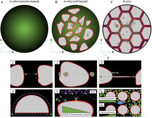
Comparison between in vitro and in vivo sample analysis. a Solution-based assays rely on simple diffusion in an essentially homogenous solution. b Cell-based in vitro assays allow analysis of dynamics of intracellular processes such as (1) extracellular diffusion, (2) transcytosis and (3) the formation of signalling filopodia such as cytonemes. c In vivo (including three-dimensional collagen/matrigel cell-based) assays involve all aspects observed in cell-based in vitro assays and further parameters of molecular diffusion such as (4) tortuosity (hindrance of diffusion path by impermeable objects), (5) transient binding of molecules to the extracellular matrix (ECM) and the cell membrane, and (6) restrictive clearance for gradient formation and the additional Z-axis that introduces further dimensions for diffusion

mRNA injection time determines distribution of fluorophore. a Microinjection at very early embryo stage (1–4 cell) generates homogenous expression of fluorophore across entire embryo, while later stage injections (16–32 cell) generates confined/mosaic patterning of fluorophore. b Depending on time of injection, patterning on embryo can be imaged with FCS or with FCCS using two or more fluorophores

Acknowledgments
This image is the copyrighted work of the attributed author or publisher, and ZFIN has permission only to display this image to its users. Additional permissions should be obtained from the applicable author or publisher of the image. Full text @ Histochem. Cell Biol.