
- Title
-
Mitf-family transcription factor function is required within cranial neural crest cells to promote choroid fissure closure
- Authors
- Sinagoga, K.L., Larimer-Picciani, A.M., George, S.M., Spencer, S.A., Lister, J.A., Gross, J.M.
- Source
- Full text @ Development

Zebrafish mitfa;tfec mutants phenocopy human colobomas. (A) At 4 dpf, mitfa−/−;tfec−/− mutants possess colobomas (outlined in red), with a range of severity (B). (C) At 48 hpf, serial sections through the distal (D) to proximal (P) axis of the CF reveal persistent expression of laminin in the mitfa−/−;tfec−/− mutant CF. The boxes indicate the areas enlarged in the insets showing laminin staining within the CF. Dorsal is upwards in all images. Scale bars: 50 µm. |

mitfa−/−;tfec−/− mutants display RPE and cNCC phenotypes. (A) At 48 hpf, mitf−/−;tfec+−/− and mitfa−/−;tfec−/− mutants display mild to severe hypopigmentation, respectively. The RPE eventually becomes normally pigmented by 4 dpf in both genotypes. (B) At 24 hpf, fewer cNCCs are present around the eye of mitfa−/−;tfec−/−;mitfa:GFP embryos than in the control (1) (***P=0.0007). However, mitfa−/−;tfec−/− mutants contain cNCCs posterior to the optic field (2). (C) Serial sections of mitfa−/−;tfec+/+;mitfa:GFP and mitfa−/−;tfec−/−;mitfa:GFP embryos at 48 hpf demonstrate that mitfa−/−;tfec−/− mutants possess fewer cNCCs in the POM surrounding the eyes. Dorsal is upwards in the images in B,C. Data are mean±s.e.m. Scale bars: 100 μm in A; 50 μm in B,C. |
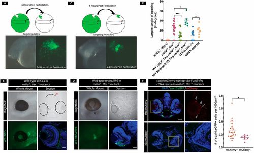
mitfa and tfec are required within cNCCs to promote CF closure. (A) NCC transplantations. At 6 hpf, wild-type dextran-labeled cells are transplanted into the cNCC-fated region of a mitfa−/−;tfec−/− embryo. At 24 hpf, transplanted cells are detected in the cNCC population. (B) Example of 4 dpf wild-type (WT) cNCC transplanted mitfa−/−;tfec−/− embryo in which the coloboma has been rescued. Transplanted cNCCs are detected in and around the eye and rescue cNCC-derived pigmented melanocytes (red arrow) confirming successful transplantation. Scale bar: 25 µm. (C) Retina/RPE transplants. At 6 hpf, wild-type dextran-labeled cells are transplanted into the RPE/retinal-fated region of a mitfa−/−;tfec−/− mutant. At 24 hpf, transplanted cells are detected within the eye, indicating successful transplantation. (D) At 4 dpf, transplanted embryos still possess colobomas. Arrow indicates a coloboma in the retina/RPE transplanted eye. (E) Quantification of colobomas (largest angle of CF opening) in rescue experiments. CF opening is significantly reduced in mitfa−/−;tfec−/− embryos transplanted with wild-type cNCCs (P=0.0004, n=14 eyes). However, colobomas are not resolved in embryos transplanted with wild-type retina/RPE cells and are actually larger than in non-transplanted control mitfa−/−;tfec−/− mutants (P=0.037, n=6 eyes). tfec cDNA injection also rescues colobomas (P=0.033, n=14 eyes). (F) pDestTol2pA2-sox10:mCherry-nostop-t2A-FLAG-tfec-pA injection reduces CF opening in mitfa−/−;tfec−/− mutants. Arrows indicate mCherry+ (rescued) neural crest cells. Quantification of sox10:eGFP+ cells shows rescue of cNCCs within the eye field (P=0.023, n=20 mCherry+, n=8 mCherry− eyes). *P<0.05, ***P<0.001. Dorsal is upwards in all images. Data are mean±s.e.m. Scale bars: 25 μm (B,D); 50 μm (F). |

tfec-/- single mutants do not possess colobomas. Whole mount and transverse sections of 4dpf tfec -/- mutants reveals mild microphthalmia and pigmentation defects. No colobomas are detected. Dorsal is up in all images. Scale bar = 100μm. PHENOTYPE:
|

Loss of cNCCs in the POM is not the result cell death. A) Whole-mount images of mitfa -/-;tfec +/- and mitfa -/-;tfec -/- embryos reveals persistent loss of cNCC within the POM at 48hpf and 72hpf. B) Quantification of TUNEL+ cNCCs. While a trend of increased cell death is present, loss of cNCC numbers cannot be attributed solely to apoptosis at 24hpf (p=0.512, n=5 embryos). |
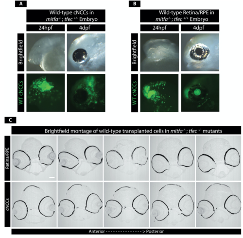
Transplantation of wild-type cells does not affect eye development. A)Wild-type cells transplanted into the region of cNCC origin in a mitfa -/-;tfec +/- embryo shows no effect on eye development. B) Similarly, transplanted wild-types cells targeting the retina/RPE in mitfa -/-;tfec +/+ embryos show no effect on eye development. C) Serial sections of representative retina/RPE and cNCC transplanted embryos. CF closure phenotypes are only detected in retina/RPE cell transplanted embryos. Dorsal is up in all images. Scale bar = 100μm. |