Duplicated lhfpl5 genes in teleost fish. (A) Phylogenic tree of representative teleost and vertebrate Lhfpl5 protein sequences. See Supplementary Table S1 for species’ names and protein accession numbers. (B) Sequence alignment of Lhfpl5 proteins from zebrafish (Danio rerio, Dr), chicken (Gallus gallus, Gg), mouse (Mus musculus, Mm), and humans (Homo sapiens, Hs). Regions of the proteins coded for by exons 1 and 3 are shaded gray. Locations of the transmembrane (TM) helices 1–4 are shown in the linear protein structure diagram. The lhfpl5atm290d and lhfpl5bvo35 mutations are indicated by the red triangle and red square, respectively.

Zebrafish lhfpl5a and lhfpl5b genes are expressed in distinct populations of sensory hair cells. Whole mount mRNA in situ hybridization for lhfpl5a(A,C,E,G,I) and lhfpl5b(B,D,F,H,J) at 1, 2, and 5 days post-fertilization (dpf). (G–J) Are details from 5 dpf larvae. NMs, neuromasts; HC, hair cell; SC, support cell. Scale bars: (A,B) = 50 μm; (C–F) = 100 μm; (G–J) = 25 μm.

lhfpl5a is required for auditory and vestibular hair cell function. (A) Boxplot showing the first peak amplitude (μV) of microphonic recordings from the inner ear of 3 dpf wild type, lhfpl5atm290d, and lhfpl5bvo35 larvae. The boxes cover the inter-quartile range (IQR), and the whiskers represent the minimum and maximum datapoints within 1.5 times the IQR. Values from individual larvae are indicated by the black dots and outliers indicated by a diamond. Asterisks indicate p < 0.01 (∗∗) by Welch’s t-test. ns, not significant. (B) Boxplot of the acoustic startle response in 6 dpf wild type and lhfpl5atm290d mutants, with or without the GFP-lhfpl5a vo23Tg transgene. Values from individual larvae are indicated by the black dots and outliers indicated by a diamond. Asterisks indicate p < 0.001 (∗∗∗) by Welch’s t-test. ns, not significant. (C–F’) FM 4–64 dye labeling assay for MET channel activity of inner ear hair cells from 6 dpf larvae [C – Wild type (+/+ or±), Dlhfpl5atm290d mutants, E – Wild type GFP-lhfpl5a vo23Tg, F – GFP-lhfpl5a vo23Tg; lhfpl5atm290]. (E’,F’) Are images of GFP-Lhfpl5a protein in the bundle of the hair cells shown above in (E,F). n = 2, 3, 12, and 4 for the genotypes in (C–F), respectively. Scale bars = 5 μm and apply to all images.

lhfpl5b is required for lateral line hair cell function. (A–C) Representative images of 5 dpf zebrafish larvae (A – Wild type; Blhfpl5atm290d; Clhfpl5bvo35) labeled with the MET channel-permeant dye FM 1–43. (D–G) Representative images of individual neuromasts from 2 and 5 dpf wild type and lhfpl5bvo35 larvae labeled with FM 1–43. Dashed lines outline the cluster of hair cells in each neuromast. (H) Quantification of normalized FM 1–43 fluorescence intensity per hair cell of 2 and 5 dpf neuromasts (n = 10 WT, 14 lhfpl5bvo35 NMs at 2 dpf; n = 6 WT, 13 lhfpl5bvo35 NMs each genotype at 5 dpf). The box plots cover the inter-quartile range (IQR), and the whiskers represent the minimum and maximum datapoints within 1.5 times the IQR. Asterisks indicate p < 0.001 (∗∗∗) by Welch’s t-test. (I,J) Rescue of FM dye labeling in lhfpl5bvo35 mutants (n = 7) by the GFP-lhfpl5a (vo23Tg) transgene. The GFP-Lhfpl5a bundle and FM 4–64 images are from the same NM for each genotype. (K) Quantification of hair cell number in L1, MI1, and O2 neuromasts from 5 dpf lhfpl5bvo35 mutants (n = 11) and wild-type siblings (n = 11). The box plots are the same as in (H). Asterisks indicate p < 0.001 (∗∗∗) by Welch’s t-test. Scale bars = 5 μm, applies to (D–G,I,J); 2 μm in (I,J) insets.

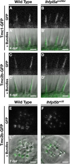
Tmc proteins do not require Lhfpl5a or Lhfpl5b for localization to the stereocilia of zebrafish hair cells. (A–D’) Representative images of Tmc1-GFP (vo27Tg) or Tmc2b-GFP (vo28Tg) in the lateral cristae of wild type (A,A’,C,C’) and lhfpl5atm290d(B,B’,D,D’) larvae. The GFP-only channel is shown in (A–D) and overlaid with a light image of the bundles in (A’–D’). (E–F’) Representative images of Tmc2b-GFP (vo28Tg) in the neuromasts of wild type (E,E’) and lhfpl5bvo35(F,F’) larvae. The GFP-only channel is shown in (E,F) and overlaid with a light image of the bundles in (E’,F’). Scale bars = 3 μm in all panels.

Lhfpl5a requires MET complex proteins Pcdh15a, Cdh23, and Myo7a for normal localization in the stereocilia of zebrafish hair cells. (A,B) Immunostain of Pcdh15a (magenta) in the lateral cristae of wild type and lhfpl5atm290d mutants at 3 dpf. Phalloidin-stained actin of the hair bundle is shown in green. Arrows indicate areas of Pcdh15a accumulation. (C–F’) Representative images of GFP-Lhfpl5a (vo23Tg) in the lateral cristae hair bundles of wild type (C,C’) and pcdh15apsi7(D,D’), cdh23nl9(E,E’), and myo7aaty220(F,F’) mutants. White arrows indicate GFP signal in the presumptive kinocilial linkages, yellow arrow heads indicate GFP signal in the stereocilia or the base of the hair bundle, and brackets indicate GFP signal in the kinocilium. Scale bars = 3 μm in (A,B); 2 μm in (C–F’).

Acknowledgments
This image is the copyrighted work of the attributed author or publisher, and ZFIN has permission only to display this image to its users. Additional permissions should be obtained from the applicable author or publisher of the image. Full text @ Front. Mol. Neurosci.