Study design. Male and female zebrafish (4–6 months old) were treated with vehicle (control, CON) or 17β-estradiol (E2, 0.1 μM). Cardiac function was assessed by echocardiography at 0, 4, 7 and 14 days after treatment initiation (n = 5/group).

Echocardiographic analysis of male and female zebrafish treated with or without E2. (A) Heart rate, (B) ejection fraction, (C) stroke volume, and (D) cardiac output 14 days after treatment initiation. Data are presented in scatter dot plots including mean ± SD. (E) Ejection fraction assessed at 4, 7 and 14 days after treatment initiation. M: male; F: female; CON: control; E2: 17β-estradiol. Data present mean ± SEM; n = 5/group; # ANOVA interaction p ≤ 0.05; ## ANOVA interaction p ≤ 0.01; * p ≤ 0.05; ** p ≤ 0.01; *** p ≤ 0.001

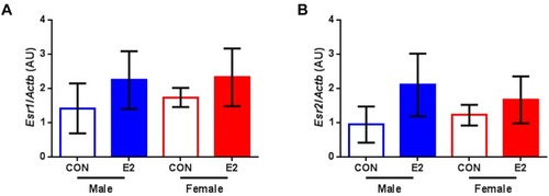
Gene expression analysis of estrogen receptors. (A) Relative mRNA levels of estrogen receptor alpha (Esr1) and (B) estrogen receptor beta (Esr2) in hearts of male and female zebrafish treated with vehicle (control, CON) or 17β-estradiol (E2) normalized to beta actin (Actb). Data present mean ± SD; n = 3/group; each replicate was derived from 2–5 pooled hearts; AU: arbitrary unit.

Acknowledgments
This image is the copyrighted work of the attributed author or publisher, and ZFIN has permission only to display this image to its users. Additional permissions should be obtained from the applicable author or publisher of the image. Full text @ Int. J. Mol. Sci.