
- Title
-
Functional analysis of new human Bardet-Biedl syndrome loci specific variants in the zebrafish model
- Authors
- Castro-Sánchez, S., Suarez-Bregua, P., Novas, R., Álvarez-Satta, M., Badano, J.L., Rotllant, J., Valverde, D.
- Source
- Full text @ Sci. Rep.

Phenotypes of zebrafish embryos at 8–12 ss, after whole mount PHENOTYPE:
|
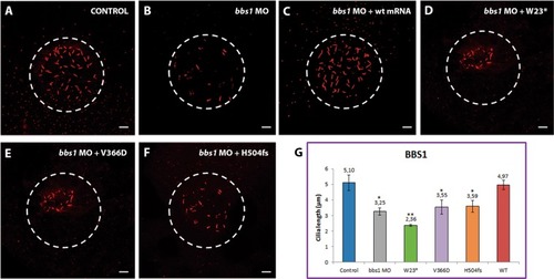
Representative images of KVs and comparison of average cilia length corresponding to PHENOTYPE:
|
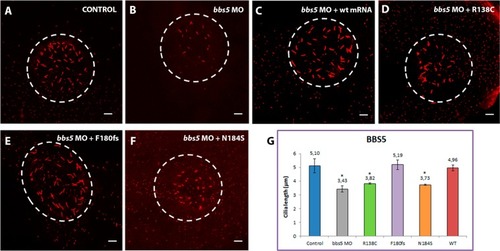
Representative images of KVs and comparison of average cilia length corresponding to PHENOTYPE:
|

Representative images of KVs and comparison of average cilia length corresponding to PHENOTYPE:
|

ZFIN is incorporating published figure images and captions as part of an ongoing project. Figures from some publications have not yet been curated, or are not available for display because of copyright restrictions. PHENOTYPE:
|