FIGURE SUMMARY
Title

Skeletal mineralisation in association with type X collagen expression is an ancestral feature for jawed vertebrates

Authors
Debiais-Thibaud, M., Simion, P., Ventéo, S., Muñoz, D., Marcellini, S., Mazan, S., Haitina, T.
Source
Full text @ Mol Bio Evol

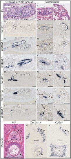
Histology and in situ hybridization on cryosections of catshark Scyliorhinus canicula skeletal tissues at stage 33. (A, B, C, V) Hematoxylin–Eosin–Saffron (HES) histological staining. (DU) Expression of Col10a1 duplicates in developing teeth and scales of catshark detected with probes specific for each duplicate. Dotted lines indicate the mesenchymal compartment limits. Asterisk marks the mineralizing matrix, often seen as empty space due to decalcification. (W, X) Expression of Col10a1.4 duplicate in comparison to Col2a1 in mineralizing vertebral tissues of catshark. Black arrowhead in the neural arch points to the site of globular mineralization; open arrowhead points to the site of lamellar mineralization; black arrowhead in the vertebral body points to the site of fibrous mineralization. Scale bars: 50 µm (AU), 200 µm (VX). Abbreviations: mat. amel., maturation stage ameloblasts; M’s, Meckel’s cartilage; notoch, notochord; od., odontoblasts; sec. amel., secretory ameloblasts.

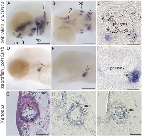
Expression of zebrafish col10a1a and col10a1b and Xenopus Col10a1 during tooth development detected with in situ hybridization. (A, B) col10a1a expression in whole-mount zebrafish larvae at 4 and 6 days post fertilization (dpf). (C) col10a1a expression in post in situ paraffin sections of zebrafish larvae at 5 dpf. (D, E) col10a1b expression in whole-mount zebrafish larvae at 4 and 6 dpf. (F) col10a1b expression in post in situ paraffin sections of zebrafish larvae at 4 dpf. (G) HE staining of a longitudinal section through a developing tooth bud of a Xenopus larva at stage NF57. (H, I) Col10a1 expression in Xenopus tooth bud on successive transversal sections. Open arrowheads point at developing pharyngeal teeth and asterisk marks the mineralizing matrix. Scale bars: 100 µm (A, B, D, E), 50 µm (C, F), 20 µm (G, H, I). Abbreviations: amel., ameloblasts; cl, cleithrum; d, dentary; m, maxilla; od., odontoblasts; op, opercle; p, parasphenoid; sec. amel., secretory ameloblasts.

Acknowledgments
This image is the copyrighted work of the attributed author or publisher, and ZFIN has permission only to display this image to its users. Additional permissions should be obtained from the applicable author or publisher of the image. Full text @ Mol Bio Evol