
- Title
-
Glucocorticoid receptor-dependent induction of cripto-1 (one-eyed pinhead) inhibits zebrafish caudal fin regeneration
- Authors
- Garland, M.A., Sengupta, S., Mathew, L.K., Truong, L., de Jong, E., Piersma, A.H., La Du, J., Tanguay, R.L.
- Source
- Full text @ Toxicol Rep
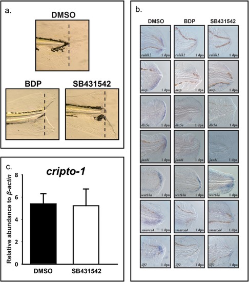
BDP and SB431542 impact larval regeneration. a) Caudal fins of 2 dpf (days post-fertilization) larvae were amputated and exposed to vehicle (0.1% DMSO), 1 μM beclomethasone dipropionate (BDP), or 100 μM SB431542. Regenerative progression was evaluated and pictures were taken at 3 dpa (days post-amputation). b) In situ localization of dlx5a, junbl, wnt10a, ilf2, smarca4, raldh2 and mvp in larvae exposed to BDP and SB431542 demonstrated a similar expression pattern. c) Exposure to SB431542 did not significantly alter expression of cripto-1. |

Partial antisense repression of Cripto-1 rescues inhibition of regeneration by BDP. a) Translation-blocking cripto-1 morpholino oligo (MO) transiently knocked down Cripto-1. The morphants developed the characteristic one-eyed pinhead phenotype by 2 days post-fertilization (dpf). b) The control and cripto-1morphants were exposed to DMSO or beclomethasone dipropionate (BDP) at 2 dpf following amputation. The dotted lines mark the plane of amputation. Regenerative progression was evaluated and pictures were taken at 3 days post-amputation (dpa). In each replicated experiment approximately 80% of the cripto-1morphants were resistant to inhibition of regeneration by BDP exposure. |

RA exposure suppress cripto-1 expression in the fin tissue and rescues BDP impaired regeneration. a) Caudal fin of 2 days post-fertilization (dpf) embryos were amputated and exposed to DMSO or either 0.01 μM or 0.1 μM retinoic acid (RA). The abundance of cripto-1was estimated in the regenerating fin tissues. The expression of cripto-1 was significantly reduced in a concentration-dependent manner compared to the control. The respective values represent the mean ± SEM and the asterisks indicate statistical significance (One-way ANOVA, n = 3, p < 0.05). b.) 2 dpf larvae were exposed to 0.1 μM RA for 8 h followed by amputation and co-exposure with 0.01 μM RA and DMSO or beclomethasone dipropionate (BDP). The abundance of cripto-1 was evaluated at 1 day post-amputation (dpa) in the regenerating fin tissue by qRT-PCR. Co-exposure with RA significantly suppressed cripto-1 expression compared to BDP exposure. The respective values represent the mean ± SEM and the asterisks indicate statistical significance (One-way ANOVA, n = 3). c). Amputated 2 dpf larvae pre-exposed with 0.1 μM RA were exposed to DMSO, BDP or co exposed with BDP and 0.01 μM RA. Regenerative progression was monitored and pictures were taken at 3 dpa. In each replicated experiment regeneration was restored in approximately 75% embryos co exposed to BDP and RA compared to BDP alone. d) In situ localization of dlx5aand junbl in the regenerating fin tissue at 1 dpa demonstrated restoration of regeneration markers in the caudal fin of larvae co-exposed with BDP and RA. |