
- Title
-
Characterization of paralogous uncx transcription factor encoding genes in zebrafish
- Authors
- Nittoli, V., Fortunato, A.E., Fasano, G., Coppola, U., Gentile, A., Maiella, S., Langellotto, F., Porreca, I., De Paolo, R., Marino, R., Fiengo, M., Donizetti, A., Aniello, F., Kondo, T., Ristoratore, F., Canzoniero, L.M.T., Duboule, D., Wilson, S.W., Sordino, P.
- Source
- Full text @ Gene X
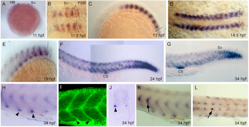
Expression of EXPRESSION / LABELING:
|
Non-somitic expression of |

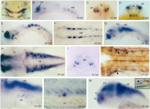
Expression of EXPRESSION / LABELING:
|

Expression of EXPRESSION / LABELING:
|

Regulation of EXPRESSION / LABELING:
PHENOTYPE:
|

Regulation of EXPRESSION / LABELING:
PHENOTYPE:
|

Unillustrated author statements |