
- Title
-
S-Sulfocysteine Induces Seizure-Like Behaviors in Zebrafish
- Authors
- Plate, J., Sassen, W.A., Hassan, A.H., Lehne, F., Köster, R.W., Kruse, T.
- Source
- Full text @ Front Pharmacol

ZFIN is incorporating published figure images and captions as part of an ongoing project. Figures from some publications have not yet been curated, or are not available for display because of copyright restrictions. PHENOTYPE:
|
|
ZFIN is incorporating published figure images and captions as part of an ongoing project. Figures from some publications have not yet been curated, or are not available for display because of copyright restrictions. PHENOTYPE:
|

DASPEI staining of 3 dpf zebrafish larve Toablate hair cells of the lateral line, 3 dpf larve were treated with 400 um neomycin for 60 min. As control untreated 3 dpf larve were used. In larve treated with neomycin, the hair cells were ablated as documented by missing fluorescence upon DASPEI staining. Control larve showed fluorescencein the hair cells of the lateral line, documenting functionality of the method. Exposure times are above the images. |
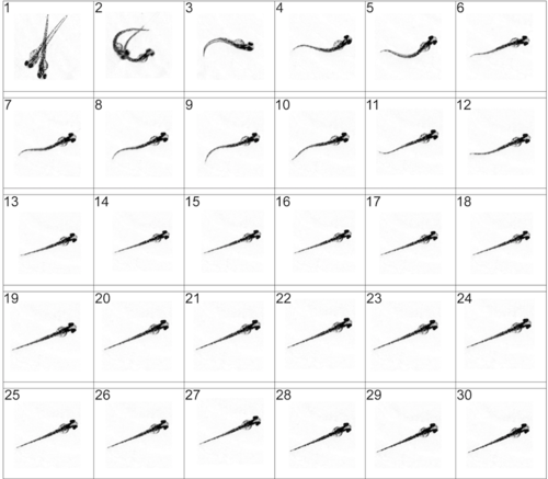
Analysis of the motor behavior of non-treated larve. Frames 3-8 have been analyzed in Supplementary figure 4. |
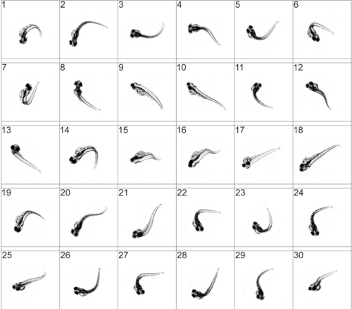
Analysis of the motor behavior of S-Sulfocysteine-treated larve. Frames 4-9 have been analyzed in Supplementary figure 4. PHENOTYPE:
|

Qualitative description of S-Sulfocysteine induced larval movement Movie data shown in supplementary figures S2 and S3 were inspected frame by frame. Representative phenotypes identified are shown for non-treated larve and larve treated with 2 mM S-Sulfocysteine (SSC). The bending of the body axis is illustrated beneath each picture, with the insets within the pictures. |

Analysis of S-Sulfocysteine long term effects on zebrafish larve (A) Acridine orange (AO) Staining of 3 dpf zebrafish larve previously treated with 2 mM S-Sulfocysteine (SSC) for 10 min, 30 min, 60 min and untreated control (if not stated otherwise: scalebar 500 um). Representative images are shown.The exposure times used are given in ms for each of the data sets. The untreated larve and larve treated with 2mM SSC for 10 min display auto fluorescence of the yolk and AO staining in regions where apoptosis is part of normal development like the olfactory placode (Van Ham et al., 2010). Larve treated for 30 min with 2mM SSC shoe intense AO staining in the brain. After 60 min of SCC treatment cell death is visualized throughout the whole larve specifying the forebrain (fb) midbrain (mb) and hindbrain (hb) (scalebar 250 um). (B) The head diameter of non treated larve and SSC treated larve were determined. The average head diameter of non treated larve was 423 um +/- 3.68 (=100%). The average head diameter of 2 mM SSC treated larve was found to be significantly higher (136.2 +/- 4.34%). Error bars indicate the standard error of the mean. The results are signifigant with p<0.01. PHENOTYPE:
|