FIGURE SUMMARY
Title

Von Willebrand Factor Mediates Pneumococcal Aggregation and Adhesion in Blood Flow

Authors
Jagau, H., Behrens, I.K., Lahme, K., Lorz, G., Köster, R.W., Schneppenheim, R., Obser, T., Brehm, M.A., König, G., Kohler, T.P., Rohde, M., Frank, R., Tegge, W., Fulde, M., Hammerschmidt, S., Steinert, M., Bergmann, S.
Source
Full text @ Front Microbiol

Streptococcus pneumoniae binds human plasma VWF. (A) Binding analyses with radioactive labeled VWF and seven S. pneumoniae strains: R6, R800 and the serotypes (st) 35A, 4 (TIGER4), 2 (D39), 23F and 12F. VWF binding was expressed as percentage of total radioactivity added. Amount of capsular polysaccharides (CPS) was categorized according to the CPS-diameter determined by electron microscopic visualization: low encapsulation <0.03μm, middle encapsulation in a range of 0.03–0.5μm and high encapsulation >0.5μm. Scale bars represent 0.5 μm in the overview figures and 0.1 μm in magnified sections. (B) Immunofluorescence visualization of VWF binding to RFP-expressing pneumococci with FITC-conjugated VWF-specific antibodies after embedding of VWF-incubated pneumococci in agarose. RFP-expressing bacteria appear in red at 540/590 nm and VWF-detection is visualized in green at 470/515 nm. VWF coated bacteria appear in yellow in the merged channel. The insets visualize a representative fluorescence signal at 3-fold magnification. Antibody controls revealed no unspecific VWF-signal detection on the surface of pneumococci (Ctrl). Scale bar represents 10 μm. Fluorescence intensity determination visualizes the VWF-bacterial colocalization within the region of interest (ROI 01).

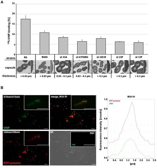
Pneumococci bind to VWF strings generated in continuous flow. (A) HMW VWF fiber generation was microscopically quantified after exposing confluently grown HUVEC to shear stress using a microfluidic device (ibidi®) at 10 dyne/cm2. FITC-conjugated VWF-specific antibodies detected HMW VWF strings. White arrows point to red fluorescent pneumococci attached to long VWF strings. (B) Attachment of RFP-expressing pneumococci to green fluorescent VWF strings was microscopically observed after 30 min in constant flow (white arrows) and was confirmed by software-based evaluation of fluorescence intensities of the defined ROI. Pictures were taken in real time using the fluorescence equipment of a confocal laser scanning microscope (SP8, Leica). Scale bar represents 10 μm.

Colocalization of pneumococci with VWF in vascular system of Danio rerio. (A) Colocalization of red fluorescent pneumococci with zebrafish-derived VWF was microscopically visualized after fixation, embedding, and staining of wild type larvae with VWF-specific rabbit FITC-conjugated antibodies and pneumococcus specific rabbit antibodies followed by goat Alexa 568-conjugated antibodies. Visualization of three representative larvae (no. 1, 2, and 3) is shown after digital zoom in magnification of the region marked by white and subsequently by black squares. All larvae are displayed from ventral side. The white square encircles intersegmental vessels of the trunk muscles in larva 1, the heart vasculature of larva no. 2 and the vessels of the gill arches of larva no. 3. White arrows of R1–R5 point to VWF colocalization with single diplococci or short pneumococcus chains appearing as yellow overlay. Colocalizations within selected regions of interest (zoom in ROI) are confirmed by corresponding overlay histograms. Scale bars represent 10 μm. (B) Visualization of RFP-expressing serotype 35A pneumococci in zebrafish larvae without and after pre-incubation of pneumococci with human VWF prior to injection of 600 cfu into the heart chamber. The red square in the zebrafish illustration depicts the region of the zebrafish tail, which is focused for visualization after immunostaining. Using a transgenic zebrafish larva, which expresses red-fluorescing endothelium (FLK1:mCherryCAAX), the position of the main blood vessel and its branching circular microvasculature within the tail region are visualized in b. After 5 h of infection with non-VWF-pre-incubated RFP-expressing pneumococci (without VWF), only single diplococci are detected within the intersegmental blood vessels of the tail region of cross linked larvae after immune staining of VWF (green) (without VWF, merge, white arrows). The yellow appearance in the merged illustration points to the recruitment of zebrafish-derived VWF to the surface of the bacteria. After 5 h of infection with VWF pre-incubated pneumococci, several bacterial aggregates were detected after immunostaining of VWF in green and bacteria in red as yellow clumps of up to 10 μm in size in the vasculature of the zebrafish tail (+VWF, merge and BF + merge, yellow arrows). (C) A further representative example of VWF-mediated pneumococcal aggregate formation with the vasculature of an infected zebrafish larva. A zoom in into the zebrafish region in close proximity to the larval heart (white square) visualizes a cluster of pneumococci (+VWF, merge, red arrows). A histogram of fluorescence signal overlaps in the aggregates is added in Figure S3B. Scale bars represent 10 μm.

Acknowledgments
This image is the copyrighted work of the attributed author or publisher, and ZFIN has permission only to display this image to its users. Additional permissions should be obtained from the applicable author or publisher of the image. Full text @ Front Microbiol