
- Title
-
Stable transgenesis in Astyanax mexicanus using the Tol2 transposase system
- Authors
- Stahl, B.A., Peuß, R., McDole, B., Kenzior, A., Jaggard, J.B., Gaudenz, K., Krishnan, J., McGaugh, S.E., Duboue, E.R., Keene, A.C., Rohner, N.
- Source
- Full text @ Dev. Dyn.
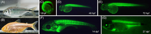
Ubi‐GFP stable transgenic line expresses GFP at different developmental stages of Astyanax mexicanus. Surface fish (A) and cavefish (B) forms of Astyanax mexicanus are a versatile model system for evolutionary and developmental biology research. C‐G, GFP expression in different developmental stages of surface fish of transgenic A. mexicanus: 18 hpf (C), 48 hpf (D), 72 hpf (E), 14 dpf (F), 27 dpf (G). hpf = hr postfertilization, dpf = days postfertilization (images not to scale)
|

Ubiquitin promoter driven transgene expresses GFP in different tissues. A, transverse section of a 14‐dpf‐old Ubi‐GFP transgenic surface fish. Inset picture indicates the location of the section. B, GFP expression in different organs of adult transgenic fish, from top to bottom: brain, liver, eyes, heart. C, Fluorescence activated cell sorting of head kidney cells from Ubi‐GFP transgenic A. mexicanus. Four distinct hematopoietic cell populations can be identified by forward scatter and side scatter: (i) myelomonocytes, (ii) progenitors, (iii) lymphocytes, and (iv) erythrocytes. Green indicates GFP signal from Ubi‐GFP transgenes, while dark grey indicates GFP signal from wild‐type control
|

A‐R, Expression of Cntnap2a‐mCherry across development. Visualization of Cntnap2a‐mCherry expression during early development (2‐28 dpf) in whole body (left column) and dorsal head view (right column). Cntnap2a expression is evident throughout the lateral line and hindbrain regions
|

Cntnap2a colocalizes with DASPEI neuromast stain. Whole body view (A‐A′′′) and zoom (B‐B′′′) show that Cntnap2a‐mCherry (red) and neuromasts of lateral line (DASPEI, green) colocalize (merge, B′′′). Dorsal head view (C‐C′′′) and zoom (D‐D′′′) demonstrates colocalization of neuromasts and Cntnap2a labeled neurons that innervate the brain (merge, D′′′). E, Sagittal aspect. F, Dorsal aspect (SB = 100 μm). Spine (Sp, purple), rhombencephalon (Rbh, pink), mesencephalon (Mes, blue), diencephalon (Dien, yellow), telencephalon (Tel, green), Cntnap2a:mCherry (red). E, Arrow, medial octavolateralis nucleus
|