
- Title
-
Abundance of Early Embryonic Primordial Germ Cells Promotes Zebrafish Female Differentiation as Revealed by Lifetime Labeling of Germline
- Authors
- Ye, D., Zhu, L., Zhang, Q., Xiong, F., Wang, H., Wang, X., He, M., Zhu, Z., Sun, Y.
- Source
- Full text @ Mar. Biotechnol.
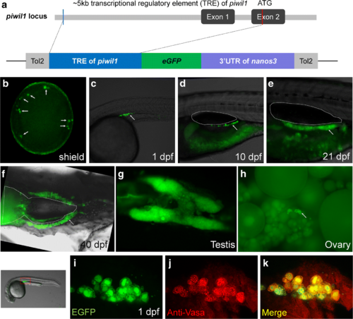
Generation and characterization of Tg(piwil1:egfp-UTRnos3)ihb327Tg. a The structure of the transcriptional regulatory region of piwil1 locus of zebrafish and the sketch map of transgenic elements. b–h Germ cell-specific expression at various developmental stages as indicated in the figure (arrows indicate the EGFP-positive germ cells; outlines indicate the swim bladder). g Dissected adult testis. h Dissected adult ovary (arrows indicate EGFP-positive oogonia or early stage of oocytes). i–k Immunofluorescence of Vasa in the transgenic embryo at 1 dpf. The selected region for imaging is indicated by a red box. iEGFP; j anti-Vasa; k merged channel
Construct:
Tg(piwil1:EGFP-UTRnanos3)
|

Using live tracing to study the correlation between the initial PGC numbers and sexual development. a Representative images of PGC-less and PGC-rich embryos at 1 dpf (a1, a5), 5 dpf (a2, a6), and 7 dpf (a3, a7). b Representative images of PGC-less and PGC-rich larva fish at 11 dpf (b1, b4), 14 dpf (b2, b5), and 20 dpf (b3, b6). c Frequency distribution of PGC number at 1 dpf; two boxes with dashed frame label the selected population of “PGC-less” and “PGC-rich.” The lateral areas of gonads were calculated and were shown at the lower right corner of images in a and b. d The sex ratio in the population of PGC-less and PGC-rich embryos. e Confocal microscopy of small and big gonads at 20 dpf with Tg(piwil1:egfp-UTRnos3)ihb327Tg transgenic fish. e1 Confocal image of small gonads. e2Higher magnification of a representative image of small gonad displaying combined channels of DAPI, F-actin, and EGFP. e3 Magnification image showing the nuclei of gonocyte in a small gonad. e4 Confocal image of big gonads. e5, e6 Higher magnification of representative images of big gonads displaying combined channels of DAPI, F-actin, and EGFP. e5 shows an example in which the germ cells have an irregular shape of nuclei; e6shows an example in which chromatin nucleolus-stage oocytes exist. e7 Magnification image showing cells with irregular shape of nuclei. e8 Magnification image showing that the nuclei of gonocyte were similar to the one in a small gonad |

The abundance of early PGCs promotes female development. a Representative images showing PGCs in wildtype and buc-overexpressed embryos at shield stage (PGCS was visualized by WISH). b Percentage of embryos with increased PGCs or normal number of PGCs in wildtype and buc-overexpressed embryos. c Representative images showing PGCs in wildtype and buc-overexpressed embryos at 1 dpf (PGCS was visualized using Tg(piwil1:egfp-UTRnanos3)ihb327Tg). d The sex ratio in population of wildtype and buc-overexpressed embryos. e RT-qPCR showing that buc was overexpressed in Tg(kop:KalTA4-UTRnanos3,CMV:EGFP)ihb8Tg/Tg(UAS:buc-UTRnanos3)ihb120Tg double-transgenic fish (dual-Tg) and Tg(UAS:buc-UTRnanos3)ihb120Tg. f The sex ratio in the populations of sibling control (Tg(kop:KalTA4-UTRnanos3,CMV:EGFP)ihb8Tg and wildtype fish), Tg(UAS:buc-UTRnanos3)ihb120Tg and double transgenics |