
- Title
-
Photoreceptor Progenitors Depend Upon Coordination of gdf6a, thrβ, and tbx2b to Generate Precise Populations of Cone Photoreceptor Subtypes
- Authors
- DuVal, M.G., Allison, W.T.
- Source
- Full text @ Invest. Ophthalmol. Vis. Sci.

gdf6a loss in tbx2b heterozygous fby carriers reduces UV cone abundance. (A) Relative abundances of UV, blue, and red cones in compound gdf6a+/s327; tbx2b+/fby in-crosses show near-complete loss of UV cones in tbx2bfby/fby mutants and partial loss of UV cones in tbx2b+/fby mutants with gdf6as327/s327 background (labeled gdf6a−/− or “−/−”). Values with matching letters are not significantly different (Kruskal-Wallis test with Mann-Whitney pairwise comparisons, gray asterisks indicate *P < 0.05, **P < 0.01, ***P < 0.001 relative to wild type (WT)/WT controls). Gdf6as327/s327 mutants have reduced blue cones and a novel reduced red cone abundance phenotype, both of which appear either unaffected or slightly enhanced by tbx2b loss. Wild-type values shown are sibling controls. (B) Representative retinal images depicting UV (magenta), blue (cyan), and red cones (green) in wild type, gdf6as327/s327, tbx2bfby/fby, and compound gdf6as327/s327; tbx2b+/fby mutants.
|
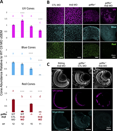
Knockdown of thrβ disrupts retinal lamination, including the photoreceptor layer, in gdf6as327/s327 mutants. (A) Thrβ knockdown with splice-blocking morpholino in gdf6as327/s327mutants (labeled gdf6a−/− or “−/−”) fails to increase UV cone abundance, instead causing near-total loss of blue and red cones. Values with matching letters are not significantly different (Kruskal-Wallis test with Mann-Whitney pairwise comparisons, gray asterisksindicate *P < 0.05, **P < 0.01, ***P < 0.001 relative to WT + control [CTL] MO controls). UV cone abundance decreased significantly, in contrast to thrβ knockdown in wildtype fish. (B) Gaps or “holes” can be seen in the photoreceptor layer of gdf6as327/s327; thrβ MO-treated whole-mount retinas (two holes indicated by asterisks in UV image). These holes are not seen in CTL MO-injected gdf6as327s327 mutants, nor in wild-type or heterozygous morphants. (C) Radial sections of 4 dpf gdf6as327/s327; thrβ MO-treated retinas show disrupted retinal lamination, with gaps in the photoreceptor layer that are occupied by cells of the inner nuclear layer, and cells disrupting the inner plexiform layer between the inner nuclear layer and ganglion cell layer (n = 9 embryos examined per group). |

A transgenic zebrafish model of conditional Thrβ disruption reveals additional roles in cone development. (A) Generation of a dominant negative thyroid hormone receptor β was accomplished via an 11-amino acid C-terminal deletion in thrβ, which was then cloned into a transgene for dominant negative thrβ (dnthrβ) and eGFP under the hsp70 promoter. Endogenous Thrβ dimerizes with other factors, such as Thrβ or RXRγ, and binds thyroid hormone to activate transcription (left), whereas dnThrβ would bind endogenous receptors but, lacking ligand and coactivator binding domains, would render dimers inactive (right). (B) Transgenic embryos express the transgene, including GFP, throughout the body. Scale bar: 1 mm. (C) Thrβ MO causes dramatic reduction in red cone abundance and increased UV abundance at 10-ng dose. Our dominant negative model shows the same increase in UV cones but also a significant increase in blue cones (HS, heat shocked). Values with matching letters are not significantly different (Kruskal-Wallis test with Mann-Whitney pairwise comparisons, gray asterisks indicate *P < 0.05, **P < 0.01, ***P < 0.001 relative to CTL MO or sibling heat-shocked [HS] controls). Thrβ MO data are normalized to CTL MO data; dnthrβ HS data are normalized to sibling HS data. |

Thrβ disruption with dnThrβ does not rescue the gdf6s327/s327 blue cone phenotype. (A) Expression of dominant negative thrβ (dnthrβ) increases UV and blue cone abundances, but in gdf6as327s327 mutants (labeled gdf6a−/− or “−/−”) the low blue cone abundance is not changed and UV cones are not increased with dnthrβ expression. Gdf6as327s327 mutants have reduced red cone abundance regardless of dnthrβ expression compared to nontransgenic wildtype siblings. Values with matching letters are not significantly different (Kruskal-Wallis test with Mann-Whitney pairwise comparisons, gray asterisks indicate *P < 0.05, **P < 0.01, ***P < 0.001 relative to WT sibling HS control). (B) Representative retinal images of wild-type sibling gdf6as327s327 and gdf6as327s327 with dnthrβ expression. |