
- Title
-
Loss of the Mia40a oxidoreductase leads to hepato-pancreatic insufficiency in zebrafish
- Authors
- Sokol, A.M., Uszczynska-Ratajczak, B., Collins, M.M., Bazala, M., Topf, U., Lundegaard, P.R., Sugunan, S., Guenther, S., Kuenne, C., Graumann, J., Chan, S.S.L., Stainier, D.Y.R., Chacinska, A.
- Source
- Full text @ PLoS Genet.
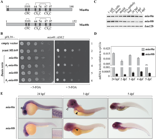
Two functional paralogues of mia40 are present in the zebrafish genome. (A) Schematics of the protein products of the two paralogues of mia40 in zebrafish. Disulfide bonds between conserved cysteines are marked. (B) The two zebrafish paralogues were expressed in yeast strains devoid of MIA40. The lethal phenotype of yeast strains depleted of MIA40 is rescued by the expression of mia40a or mia40b. Growth is restored to levels of native yeast Mia40 upon fusion of Danio rerio paralogues to the b2-mitochondria targeting presequence. (C-E) The spatiotemporal expression of mia40 paralogues in zebrafish development was monitored by RT-PCR (C), RT-qPCR (D) and in situ hybridization (E). (C) Transcripts for both mia40 paralogues are maternally contributed as well as expressed by the embryo during early zebrafish development. (D) Relative to rpl13a expression, mia40a mRNA levels are significantly higher compared to the levels of mia40b. Error bars correspond to SEM; CT values are presented in S8 Table. (E) mRNA expression patterns of mia40a and mia40b by in situ hybridization at indicated time points. At 24 hpf, mia40a expression is detected in the eye, brain (arrow), and somites, while mia40b expression is detected only in the head. Insets show a higher magnification of the somites. At 3 dpf, mia40a expression is enriched in the eye (white arrowhead) and brain (black arrows), and both mia40a and mia40b are expressed in the liver (black arrowheads). mia40a expression is greatly reduced by 5 dpf, whereas mia40b expression is strong in the head, branchial arches, epidermis, and lateral line primordia. |

Mia40a is essential for survival in zebrafish. TALENs were used to obtain mia40a and mia40b mutants (A, B). (A) A TALEN was designed to target the locus encoding Phe68 of mia40a. The identified 8 base pair (bp) deletion and the predicted protein product for the mia40abns292 allele are shown (bottom panels). In bold are TALEN arm-recognized DNA sequences. (B) A TALEN targeting the CPC-encoding region of mia40b was used to obtain a mutant with a truncated form of Mia40b. The identified 10 bp deletion leads to a frameshift mutation with a premature stop codon. The predicted protein product for the mia40bbns293 allele is shown (bottom panels). In bold are TALEN arm-recognized DNA sequences. (C) mia40a homozygous mutants do not present any gross morphological defects compared to their siblings at 5 dpf. Scale bar, 500 μm. (D) Homozygous mia40a mutants die during mid-larval to juvenile developmental stages while mia40b mutants survive to adulthood and give rise to progeny. The results are derived from AB or Tg(Xla.Eef1a1:mlsEGFP) genetic background. n: number of analysed individuals. PHENOTYPE:
|

Abnormal mitochondrial structures are found in the skeletal muscles of mia40a mutants. (A) Embryos obtained from an in-cross of heterozygous mia40a+/- siblings in the Tg(Xla.Eef1a1:mlsEGFP) background were stained with phalloidin to visualize actin filaments of the skeletal muscle at 4 dpf. Abnormal, enlarged mitochondrial structures are found in the skeletal muscles of the mia40a mutants (arrowheads). To better visualize the GFP-positive inclusions, a magnified image is shown on the side. Images are maximum projections. Scale bar, 50 μm. All images are lateral views, anterior to the left. (B) wild-type and mia40a-/- larvae in the Tg(Xla.Eef1a1:mlsEGFP) background were stained with an anti-Tomm20 antibody to mark the outer mitochondrial membrane (OMM). DNA was counterstained with DAPI. Single plane sections are shown. The arrowhead points to a GFP-positive inclusion. Scale bar, 5 μm. All images are lateral views, anterior to the left. (C) wild-type or mia40a-/- siblings in AB background were subjected to transmission electron microscopy (TEM) analysis at 5 dpf. Arrowheads point to well defined mitochondrial membranes in wild-type larvae. Abnormal membranous structures found in the mia40a mutants are magnified (inset). Scale bar, 500 nm. M: Mitochondria. |

ZFIN is incorporating published figure images and captions as part of an ongoing project. Figures from some publications have not yet been curated, or are not available for display because of copyright restrictions. PHENOTYPE:
|
|
ZFIN is incorporating published figure images and captions as part of an ongoing project. Figures from some publications have not yet been curated, or are not available for display because of copyright restrictions. |
|
ZFIN is incorporating published figure images and captions as part of an ongoing project. Figures from some publications have not yet been curated, or are not available for display because of copyright restrictions. |