
- Title
-
Conditional mutagenesis by oligonucleotide-mediated integration of loxP sites in zebrafish
- Authors
- Burg, L., Palmer, N., Kikhi, K., Miroshnik, E.S., Rueckert, H., Gaddy, E., MacPherson Cunningham, C., Mattonet, K., Lai, S.L., Marín-Juez, R., Waring, R.B., Stainier, D.Y.R., Balciunas, D.
- Source
- Full text @ PLoS Genet.

Generation and testing of a conditional (“floxed”) tbx20 allele. a. Experimental design. b. Genotyping of adult fish for intron 2 loxP site. c. Results of nested PCR screening for loxP integration into intron 1. d. Sequence of intron 1 loxP integration in recovered tpl145 floxed tbx20 allele. e. Genotyping of “F1” adults. Primer binding sites are shown as black arrows, loxP sites as red or blue triangles, exon as an open box. f, g. Induction of tbx20 loss of function by injection of Cre mRNA. f. One quarter of embryos obtained by in-crossing tbx20tpl145 heterozygotes and injected with Cre mRNA display a consistent, severe heart development defect. g. Genotyping of embryos with severe heart defects (lanes 1–3) and phenotypically normal siblings (lanes 4–8). L, GeneRuler DNA Ladder (ThermoFisher Scientific). Genotyping lanes 1 and 5 correspond to images in f. h, i. Induction of tbx20 deletion by 4-hydroxytamoxifen. tbx20tpl145 heterozygote was crossed to Tg(ubi:CreERT2) line. GFP-positive embryos were collected at 2dpf and incubated with 5μM 4-HT for 24 hours. j. Adults raised from Cre-injected tbx20tpl145/+ embryos were incrossed, resulting in approximately 1/4 of embryos (36/128, 28%) with severe heart defects. k. Confirmation of Cre-mediated excision of the second intron of tbx20 by sequence analysis. l, m. Analysis of excision efficiency by qPCR. l. Excision efficiency was assessed by qPCR in embryos treated with 4-HT at various concentrations at different time points and in two different ubb:CreERT2 driver lines. The y-axis indicates un-excised tpl145 normalized to the untreated control. m. Images of tbx20 phenotypes shown after treatment with 4-HT. Individuals 1, 2, and 3 from l correspond to images labeled 1, 2, and 3. PHENOTYPE:
|
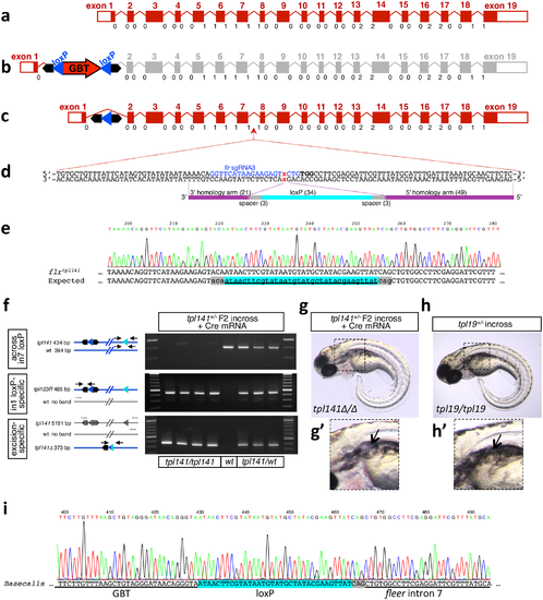
Conversion of a Cre-revertible Gene Breaking Transposon (GBT) allele to a conditional allele. a. Diagram of the fleer locus. Exons drawn to scale, introns not to scale. Below each intron-exon junction reading frame phase is indicated. b. Diagram of the fleer gene trap allele flrtpl19. c. Diagram flrtpl19R locus reverted by Cre-mediated excision of the gene trap cassette. d. Diagram of flr sgRNA3 target site and antisense oligonucleotide HDR template. e. Sequence of loxP integration into intron 7, resulting in floxed fleer allele tpl141. f, g, h. Cre-mediated excision of exons 2–7 of fleer. f. Induction of fleer deletion by Cre mRNA. g. One quarter of embryos obtained by in-crossing fleertpl141 heterozygotes and injected with Cre mRNA display a phenotype consistent with fleertpl19 homozygotes in h, including formation of kidney cysts shown in g’ and h’. i. Sequence of the excision amplicon. |

Mutagenesis of aldh1a2. a. Diagram of the aldh1a2 locus. Exons drawn to scale, introns not to scale. Below each intron-exon junction reading frame phase is indicated. b. Two highly active intronic aldh1a2 sgRNAs, aldh1a2 sgRNA1 and aldh1a2 sgRNA4, flank exon 8. Sequence corresponding to single guide RNA is shown in blue, PAM motif is in bold, and expected Cas9 cut site is indicated by a red x. c. Two independent aldh1a2 exon 8 deletion alleles recovered after co-injection of aldh1a2 sgRNA1 and aldh1a2 sgRNA4 along with nCas9n mRNA. For Sanger sequencing of the alleles, see S3 Fig. d, e. Deletion of exon 8 of aldh1a2 results in expected loss-of-function phenotype. F1 fish heterozygous for aldh1a2tpl137 and aldh1a2tpl138 deletions were crossed to each other. d. Images of 3 dpf embryos displaying wild type (top) and the expected aldh1a2 loss of function phenotypes: lack of pectoral fins, shortened hindbrain brain and cardiac edema. In addition, most of the 3 dpf embryos displaying these phenotypes had curved tails (d, bottom) consistent with uneven left/right somite numbers. e. aldh1a2tpl137/tpl138 trans-heterozygotes lack pectoral fin buds as revealed by loss of tbx18 expression at 32 hpf. f. Expression of tcf21 persists in the first and second branchial arches of aldh1a2tpl137/tpl138 trans-heterozygotes at 32 hpf. g. Diagram of the HDR template oligonucleotide used to knock in the loxP into aldh1a2 sgRNA1 target site. h. Sequencing of the precise loxP knockin allele. Additional knock-in alleles recovered from other F0 families are shown in S5 Fig. i. Genotyping of 16 phenotypically normal 3 dpf embryos from a cross between F1 fish heterozygous for aldh1a2tpl139 loxP knock-in results in Mendelian ratios of aldh1a2tpl139/tpl139 (red arrows), aldh1a2tpl139/wt (yellow arrows) and aldh1a2wt/wt embryos. PCR fragments from two of the embryos were sequenced to further confirm homozygosity for the loxP site. EXPRESSION / LABELING:
PHENOTYPE:
|

ZFIN is incorporating published figure images and captions as part of an ongoing project. Figures from some publications have not yet been curated, or are not available for display because of copyright restrictions. EXPRESSION / LABELING:
|