
- Title
-
Generation of a genetically encoded marker of rod photoreceptor outer segment growth and renewal
- Authors
- Willoughby, J.J., Jensen, A.M.
- Source
- Full text @ Biol. Open
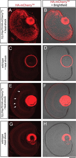
Expression of HA-mCherryTM in the eye of a 69.5 hpf larva at 1.5 hours hpHS (heat shocked at 68 hpf). (A, C, E, G) Single confocal z-section images of an eye labeled with anti-HA antibodies. (B, D, F, H) Single confocal z-section image of an eye labeled with anti-HA antibodies merged with DIC-like image. (A, B) Heat-shocked Tg(hsp70:HA-mCherryTM) larva shows ubiquitous expression of HA-mCherryTM in retinal cells. (C, D) Heat-shocked wild-type eye shows only weak autofluorescence. (E, F) In the absence of heat-shock, an eye of a Tg(hsp70:HA-mCherryTM) larva shows only weak expression of HA-mCherryTM in a small number of amacrine cells (arrows). (G, H) An eye of a wild-type larva shows weak autofluorescence in the absence of heat-shock. Scale bar: A–H, 50 µm. |
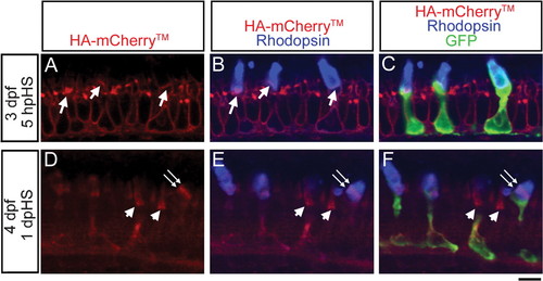
Expression of HA-mCherryTM in photoreceptors after heat-shock at 3 dpf. (A–C) At 5 hpHS, a single confocal z-section of a Tg(hsp70:HA-mCherryTM) photoreceptor layer labeled with anti-HA antibody (red), anti-Rhodopsin antibody (blue) and GFP-expressing rods (green) shows membrane expression of HA-mCherryTM in rods and neighboring cones, and a stripe of HA-mCherryTM at the base of rod outer segments (arrows, A, B). (D–F) At 1 dpHS, a single confocal z-section of a Tg(hsp70:HA-mCherryTM) photoreceptor layer labeled with anti-HA antibody (red), anti-Rhodopsin antibody (blue) and GFP-expressing rods (green) shows that membranous HA-mCherryTM labeling in the cell body and inner segment has largely disappeared and a stripe of HA-mCherryTM in a rod outer segment has been displaced distally (double arrows, D–F). Continuous HA-mCherryTM labeling largely fills cone outer segments (arrowheads, D–F). Scale bar: A–F, 5 µm. |
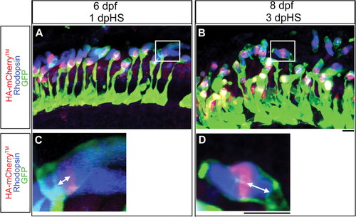
Expression of HA-mCherryTM in photoreceptors after heat-shock at 5 dpf. Confocal z-projections of Tg(hsp70:HA-mCherryTM) photoreceptor layers labeled with anti-HA antibody (red), anti-Rhodopsin antibody (blue) and with GFP-expressing rods (green). (A) At 6 dpf, 1 dpHS, HA-mCherryTM in rod outer segments is seen as a stripe, an oval or a circle, depending on the orientation of the outer segment to the plane of section. (C) A magnification of the boxed area in (A) shows the displacement of the HA-mCherryTM stripe from the base of the outer segment in 2-dimensions. (B) At 8 dpf, 3 dpHS, HA-mCherryTM in rod outer segments is seen as a stripe, an oval or a circle, depending on the orientation of the outer segment to the plane of section and HA-mCherryTM has moved distally compared to that seen at 1 dpHS. (D) A magnification of the boxed area in (B) shows the displacement of the HA-mCherryTM stripe from the base of the outer segment in 2-dimensions. Scale bars: 5 µm, A, B; 5 µm, C, D. |

Expression of HA-mCherryTM in photoreceptors after heat-shock in adult fish. Confocal z-projections of adult Tg(hsp70:HA-mCherryTM) photoreceptor layers labeled with anti-HA antibody (red), anti-Rhodopsin antibody (blue) and with GFP-expressing rods (green) at 1 dpHS (A), 4 dpHS (B), 7 dpHS (C), 9 dpHS (D), 15 pHS (E), 16 dpHS (F). Over the time-course, HA-mCherryTM moves distally toward the RPE, and has largely disappeared from rod outer segments by 16 dpHS (F). Scale bar: 20 µm, A–F. |