
- Title
-
CCAAT/enhancer-binding protein-β functions as a negative regulator of Wnt/β-catenin signaling through activation of AXIN1 gene expression
- Authors
- Park, S., Lee, M.S., Gwak, J., Choi, T.I., Lee, Y., Ju, B.G., Kim, C.H., Oh, S.
- Source
- Full text @ Cell Death Dis.
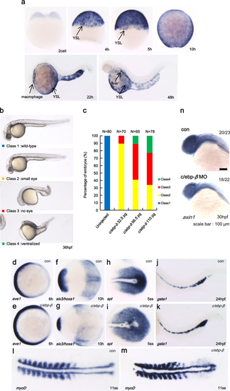
a Spatial and temporal expression of c/ebp-β in developing zebrafish embryos determined by whole-mount in situ hybridization. b Injection of zebrafish c/ebp-β mRNA induced ventralized phenotype at 36 hpf embryos. c Percentage of embryos displaying specific phenotypes following c/ebp-β mRNA injection. d–m Expression of marker genes in c/ebp-β injected embryos. eve1, a ventral marker (d, e); six3, anterior neural marker; hoxa1, posterior neural marker at the tail bud stage (f, g); spt, posterior mesoderm marker (h, i) in intermediate cell mass (ICM) in the 5-somite stage embryo; gata1 expression in ICM (j, k); myoD expression in the 11-somite stage embryo (l, m). Embryos are shown in a dorsal view with anterior to the left. cont control mRNA, 5ss 5-somatic stage, 11ss 11-somatic stage. n The expression of axin1 was examined by whole-mount in situ hybridization analysis after c/ebp-β MO injection. con control MO |
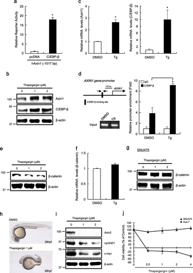
a Huh-7 cells were transfected with hAxin-1017 and C/EBP-β expression plasmid and pRL-CMV plasmid. After 48 h, luciferase activities were measured. b–f Huh-7 cells were incubated with the vehicle (DMSO) or thapsigargin (1 μM or indicated concentration) for 15 h. Whole-cell lysates prepared from Huh-7 cells were subjected to western blot analysis against the indicated antibodies (b). Real-time PCR for Axin1 and C/EBP-β were performed using total RNA prepared from Huh-7 cells (c). Chromatin samples were prepared and subsequently subjected to ChIP analysis against the anti-C/EBP-β antibody or control IgG. The amounts of immunoprecipitated C/EBP-β promoter regions were quantified by real-time PCR (d). Cytosolic proteins were subjected to western blot analysis against the β-catenin antibody (e) and total RNA was subjected to real-time PCR for β-catenin expression (f). g SNU475 cells were treated for 15 h with thapsigargin and cytosolic proteins were subsequently analyzed by western blot analysis against the β-catenin antibody. h Effect of thapsigargin on zebrafish embryonic development. i Lysates prepared from Huh-7 cells treated for 15 h with the vehicle (DMSO) or thapsigargin (1 μM) were subjected to western blot analysis against the indicated antibodies. j Effect of thapsigargin on hepatoma cell growth. In a–c, d, f, and j, results are the average of three experiments, and bars indicate standard deviations. *P < 0.05, compared with the control PHENOTYPE:
|