
- Title
-
Mutation in the intracellular chloride channel CLCC1 associated with autosomal recessive retinitis pigmentosa
- Authors
- Li, L., Jiao, X., D'Atri, I., Ono, F., Nelson, R., Chan, C.C., Nakaya, N., Ma, Z., Ma, Y., Cai, X., Zhang, L., Lin, S., Hameed, A., Chioza, B.A., Hardy, H., Arno, G., Hull, S., Khan, M.I., Fasham, J., Harlalka, G.V., Michaelides, M., Moore, A.T., Coban Akdemir, Z.H., Jhangiani, S., Lupski, J.R., Cremers, F.P.M., Qamar, R., Salman, A., Chilton, J., Self, J., Ayyagari, R., Kabir, F., Naeem, M.A., Ali, M., Akram, J., Sieving, P.A., Riazuddin, S., Baple, E.L., Riazuddin, S.A., Crosby, A.H., Hejtmancik, J.F.
- Source
- Full text @ PLoS Genet.

Relative expression of Clcc1 in mouse eye tissues at various ages, distribution of clcc1 mRNA in the zebrafish, and CLCC1 protein in the human retina. (a) Expression of Clcc1 mRNA in the cornea, lens, iris, optic nerve, and retina by qRT-PCR at different ages. Values represent the mean (± SD) on an arbitrary scale (y axis) and were calculated from at least three independent experiments. While Clcc1 is expressed in all tissues tested, ocular expression is greatest in the retina and least in the lens. (b-f) In situ hybridization of clcc1 probes in zebrafish. clcc1 Is expressed widely in zebrafish. Staining with a digoxigenin-labeled cRNA probe shows a strong signal (black arrows) in the hindbrain (HB), swim bladder (SB), and eye at 1 dpf (b, c), and in the ganglion cell layer (GCL), outer nuclear layer (ONL), and retinal pigmented epithelium (RPE) at 3 dpf (d, e and f); OS. Scale Bar: 100 μm. (g–j) IHC of formalin fixed and paraffin embedded human retinal sections demonstrated CLCC1 is expressed extensively in the retina and optical nerves. High magnification (g,i) shows more intense CLCC1 staining (arrow) in the lamina cribrosa (LC), optic nerve (ON), ganglion cell layer (GCL), inner nuclear layer (INL), outer nuclear layer (ONL) and retinal pigmented epithelia (RPE) in the retina (counter stain is methyl green). Scale Bar: g, h, 50 μm; i, j, 20μm. EXPRESSION / LABELING:
|
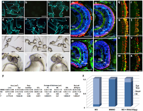
Zebrafish eye development disturbed by knockdown of clcc1 expression. Validation: Injection of the 5’-modified EGFP (a) or the unmodified EGFP (d) gave a fluorescent signal (arrowheads). Co-injection of the clcc1-MO eliminated the fluorescent signal from morpholino-sensitive 5’-modified EGFP mRNA (b) but not unmodified EGFP (e). Co-injection of the MM-MO had no effect (c, f). Eye Size: Injection of the clcc1-MO (h, k) significantly reduced eye size (black arrows) compared to MM-MO (i, l) and buffer-injected (g, j) embryos. a-f: 24 hpf, (g-l): 36 hpf. Retinal frozen sections: from 4 dpf MM-MO- (m, n) and clcc1-MO-injected (o, p) embryos stained for PKCß1 (bipolar cells, green), Zpr-1 (cone receptors, red, n and p), 1D1 (rod receptors, red, m and o), and DAPI (nuclei, blue). clcc1-MO-injected embryos show decreased thickness of ONL and IPL layers. MM-MO-injected (q, s, u, w) and clcc1-MO-injected (r, t, v, x) embryos were stained with anti-blue opsin (q, r, green), anti-green opsin (s, t, green), anti-red opsin (u, v, green), or anti-UV opsin (w, x, green), and 4D2 (all, Rhodopsin, rods, red). All photoreceptors in clcc1-MO-injected embryos show reduced staining and damaged photoreceptor cell structure, with the greatest decreases in blue and green opsin cones. m-x: 4 dpf. Scale Bar: m-p: 50 μm, q-x: 10 μm. Comparison of eye size and retinal layers: (y). GCL = ganglion cell layer, IPL = inner plexiform layer, INL = inner nuclear layer, and ONL = outer nuclear layer. Proportions of embryos with eye size phenotype: (z) with clcc1-MO injection and rescue by coinjected clcc1 WT mRNA. Lens and eye areas are given in mm2, and retinal thicknesses are given in μm. Forty clcc1-MO-treated embryos and 41 MM-MO-treated embryos were analyzed. |

Retinal morphology and function is damaged in TALEN clcc1-KO zebrafish. (a-d) Merged photographs of frozen retinal sections prepared from the heads of 5 dpf larvae. Merged photos of frozen sections from KO (b, d) and WT (a, c) embryos stained for PKCß1 (bipolar cells, green), 4D2 (rod receptors, red, a and b), Zpr-1 (cone receptors, red, c and d), and DAPI (nuclei, blue). clcc1 KO embryos show destruction of the rod photoreceptor layer (b) compared with WT (a). While somewhat better preserved than in the morpholino clcc1 knockdown embryos, cone photoreceptors and the other retinal layers also appear decreased. (e) No background, 20mM sodium aspartate, mutant cone responses are 50% depressed relative to WT. (f) PIII 6dpf Talenclcc1 vs WT. Raw means and SEMs (all spectra). Dark adapted, no background. Cone spectral sensitivity is depressed about 60% in mutants. (g) No background, 50μM CNQX. For signals from ON bipolar cells, which are 2X more sensitive than cone signals, TALEN clcc1 mutant responses decrease by over 50%. Stimuli are saturating at 490nm. (h) b2 5dpf Talenclcc1 vs WT. Raw means and SEMs (all spectra). Dark adapted, no background. ON bipolar cell spectral sensitivity is depressed over 50% in mutants. Sensitivity axis is in units of nV per quantum as calculated from the amplitude of responses to constant quanta stimulation across the spectrum (Eq 1). The quanta level of 2500 hν·μm−2·s−1 at the cornea is below semi-saturation for all cone types. (i) coinjection of zebrafish embryos with WT but not p.D25E mutant clcc1 mRNA can rescue the KO phenotype. *p 0.022 vs. WT injected with buffer control, p = 0.0082 vs KO injected with p.D25E mutant clcc1 mRNA, ** p = 0.00095 vs. WT injected with buffer control *** p > 0.00012 vs. WT injected with buffer control. |

Unillustrated author statements PHENOTYPE:
|