
- Title
-
Functions of the COPII gene paralogs SEC23A and SEC23B are interchangeable in vivo
- Authors
- Khoriaty, R., Hesketh, G.G., Bernard, A., Weyand, A.C., Mellacheruvu, D., Zhu, G., Hoenerhoff, M.J., McGee, B., Everett, L., Adams, E.J., Zhang, B., Saunders, T.L., Nesvizhskii, A.I., Klionsky, D.J., Shavit, J.A., Gingras, A.C., Ginsburg, D.
- Source
- Full text @ Proc. Natl. Acad. Sci. USA
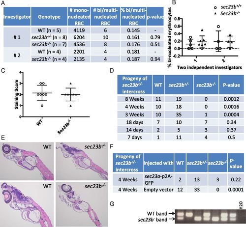
Mortality in Sec23b-deficient ZF between 18 and 21 d is rescued by Sec23a. (A and B) sec23b heterozygous (sec23b+/−) ZF with a 53-bp deletion were generated with CRISPR/Cas9. Binucleated erythrocyte counts from peripheral blood of ZF offspring from a sec23b+/− × sec23b+/− intercross were evaluated by two independent investigators blinded to the ZF genotype. (C) sec23b−/− ZF exhibit indistinguishable o-dianisidine staining compared with WT clutchmate controls. Scores 1–3 correspond to staining intensities (3 = highest staining intensity). (D) sec23b−/− ZF (generated from sec23b+/− intercrosses) die between 18 and 21 d. (E) H&E stains of sec23b−/− and WT ZF at day 16. sec23b−/− ZF appear histologically normal. (Magnification: 10×.) (F) One-cell–stage embryos generated from sec23b+/− ZF intercrosses were injected with a vector (designed to integrate in the ZF genome) that expresses Sec23a and GFP or GFP alone as control. Genotyping at 4 wk of age demonstrates rescue of sec23b−/− mortality by Sec23a. (G) PCR genotyping assay demonstrating 2 sec23b−/− ZF, expressing Sec23a from a ubiquitin promoter, alive at 4 wk of age. PHENOTYPE:
|

sec23b-/- ZF histology and GFP expression of ZF injected with a vector (designed to integrate into the ZF genome) expressing Sec23a and GFP from a ubiquitin promoter. (A) sec23b-/- ZF exhibit normal pancreas histology at day 16 and (B) normal staining with o-dianisidine at day 7 compared to WT clutchmate controls. (C) The sec23a cDNA is expressed from a ubiquitin promoter with a P2A sequence separating it from a sequence encoding GFP. (D) Ubiquitous Sec23A expression (approximated by GFP), as expected from vector integration into onecell stage embryos. PHENOTYPE:
|