
- Title
-
Triptolide attenuates proteinuria and podocyte apoptosis via inhibition of NF-κB/GADD45B
- Authors
- Wang, L., Zhang, L., Hou, Q., Zhu, X., Chen, Z., Liu, Z.
- Source
- Full text @ Sci. Rep.
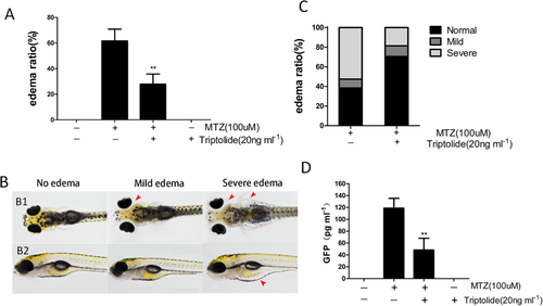
Triptolide treatment alleviated MTZ-induced oedema and proteinuria. (A,C) Quantification of the oedema ratio (n = 30) and severity of oedema. The embryos were treated with 100 μM MTZ from 84 hpf to 132 hpf. Triptolide pretreatment obviously reduced MTZ-induced oedema percentage and severity. (B) Representative figure shows the severity of oedema (arrowhead) from a dorsal view (B1) and side view (B2). (D) GFP Quantification using ELISA showing that triptolide pretreatment reduced the MTZ-induced proteinuria (n = 3). **P < 0.01. (On the basis of three triplicate tests). PHENOTYPE:
|

The effects of triptolide on podocin expression and distribution in MTZ-treated Tg(pod:gal4;UAS:NTR-mCherry) zebrafish. (A) The expression and distribution of podocin in different groups was measured and examined at 24 h, 30 h and 36 h after MTZ treatment in 84 hpf larvae; (a1–a3) normal control (CTL); (b1–b3) MTZ model group; (c1–c3) MTZ + triptolide group. (B) No obvious mean immunofluorescence intensity (GL) differences were observed between the three groups at 30 h and 36 h. (C) Positively stained area to the glomerular area (AR) revealed a remarkably reduction at 36 h in the MTZ model group, but remained strong in the triptolide treatment group. (n = 3), **P < 0.01, (original magnification, x1000). |
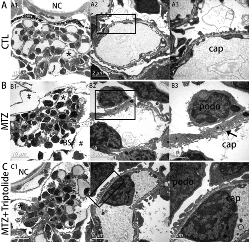
Triptolide reversed podocyte foot process effacement in MTZ-treated Tg(pod:gal4;UAS:NTR-mCherry) zebrafish. (A) Electron micrograph of normal glomerular vascular loop morphology and foot process development at 6 dpf in wild-type larvae. (B) Glomerular morphology of zebrafish treated with MTZ for 24 h. At low magnifications, an enlarged space outside of Bowman’s capsule was observed, indicating oedema (B1, indicated by #). Extensive foot process effacement was observed at higher magnifications (B3, indicated by arrows). (C) Triptolide prevented most foot process effacement. (n = 3). *Capillary that was magnified. podo, podocyte; cap, capillary space; BS, Bowman’s space; NC, notochord. Scale bar = 10 μm (A1,B1,C1), 2 μm (A2,B2,C2), 1 μm (A3,B3,C3). PHENOTYPE:
|
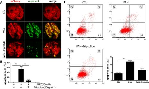
Triptolide suppressed podocyte apoptosis. (A) 84 hpf larvae treated with MTZ for 24 h exhibit remarkable podocyte apoptosis signalling compared to the untreated group (CTL) on immunostains against cleaved caspase-3. The number of apoptotic cells was significantly reduced in the triptolide treatment group. (B) Quantification of caspase-3-stained area (green) to mCherry (red). Data are expressed as the means ± SEM. The confocal images are shown at a maximum intensity projection. (n = 3), **P < 0.01; *P < 0.05. (Original magnification, x1000). (C) Annexin V-FITC/PI staining followed by FCM showed podocytes apoptosis induced by PAN was alleviated by triptolide. **P < 0.01. (On the basis of three triplicate tests). PHENOTYPE:
|

ZFIN is incorporating published figure images and captions as part of an ongoing project. Figures from some publications have not yet been curated, or are not available for display because of copyright restrictions. |
|
ZFIN is incorporating published figure images and captions as part of an ongoing project. Figures from some publications have not yet been curated, or are not available for display because of copyright restrictions. PHENOTYPE:
|