
- Title
-
Nucleo-cytoplasmic transport of TDP-43 studied in real time: impaired microglia function leads to axonal spreading of TDP-43 in degenerating motor neurons
- Authors
- Svahn, A.J., Don, E.K., Badrock, A.P., Cole, N.J., Graeber, M.B., Yerbury, J.J., Chung, R., Morsch, M.
- Source
- Full text @ Acta Neuropathol.

Fluorescent primary motor neurons in the zebrafish spinal cord at 3 dpf. a Maximum intensity projection of a z-stack of a motor neuron revealing the complete axonal arbour (yellow) projecting into and around the myotome (not shown). At the cell body the spindly dendritic arbour can also be observed. The nucleus is visualised in mCerulean3 (cyan). Traversing the image is the axon of a spinal interneuron. Scale = 10 µm. b A separate fluorescent motor neuron (bi), demonstrating the mnx1-driven membrane-bound mKO2-CAAX (yellow, bii), nuclear H2B-mCerulean3 (cyan, biii) and HsaTDP43WT-linked eGFP (green, biv). Scale = 2 µm EXPRESSION / LABELING:
|

Nuclear TDP-43 accumulations are highly motile. Fast resonance scan of a motor neuron cell body with cytoplasmic mTagBFP (magenta) and nuclear eGFP-TDP43WT (green) revealed that these TDP-43 accumulations rapidly dissociate and coalesce. The full time-lapse video gives the best example of the TDP-43 motility (Online Resource 1). Arrows indicate and follow distinct accumulations over the imaging period. Scale = 1 µm EXPRESSION / LABELING:
|

Microglial uptake of TDP-43. Sequence of a spinal microglia (mCherry-CAAX, red) migrating toward and engulfing an eGFP-TDP43WT (green)-expressing motor neuron during UV-induced degeneration. Scale = 10 µm EXPRESSION / LABELING:
|

Expansion and dissolution of a motor neuron over the course of UV-induced degeneration in the absence of microglia. ai Cytoplasmic mTagBFP signal pre-irradiation. aii–iv Cytoplasmic changes during UV-induced degeneration. bi eGFP-TDP43WT distribution in the soma of the neuron pre-irradiation. bii–iv eGFP-TDP43WT mislocalises into the cytoplasm during degeneration. c Merged images. Scale = 2 µm EXPRESSION / LABELING:
PHENOTYPE:
|

Blebbing and TDP-43 mislocalisation in a motor neuron over the course of UV-induced degeneration after depletion of microglia. ai Cytoplasmic mTagBFP signal pre-irradiation. aii eGFP-TDP43WT distribution in the soma of the neuron pre-irradiation. aiii Merged images. bi–iv eGFP-TDP43WT signal redistributes into the cytoplasm and shows the blebbing of the cell soma during degeneration. Note the apparent fragmentation of the nucleus, in contrast to the concentration of nuclear eGFP-TDP43WT in the previous figure. Scale = 2 µm EXPRESSION / LABELING:
PHENOTYPE:
|

Fragment release from a neuron undergoing UV-induced degeneration in the absence of functional microglia. ai Cytoplasmic mTagBFP signal pre-irradiation. aii eGFP-TDP43WT distribution in the soma pre-irradiation. aiii Merged images. bi–vi mTagBFP during UV-induced degeneration. Concentrated fragments can be observed separating from the external membrane before the cytoplasm disperses. ci–vi eGFP-TDP43WT during degeneration. Concentrated fragments can be observed collecting and releasing from the membrane. The concentrated (pyknotic) nuclear core gradually fragments. Scale = 2 µm |
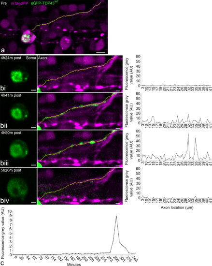
Axonal redistribution of eGFP-TDP43WT during UV-induced degeneration and microglia depletion. a Pre-irradiation, line is a projection of the axon vector used for signal analysis. Scale = 5 µm. bi–iv At each time point from left to right the three windows represents a single time point illustrating the cell body and the TDP-43 distribution (soma, eGFP-TDP43WT in green), the axonal projection (axon, mTagBFP in magenta incl. neurite vector as yellow line), and the fluorescence intensity of eGFP in the segments along the neurite vector. c Line graph demonstrating the time course of the mean eGFP fluorescence intensity along the axon post-stress induction. Scale = 2 µm. Note: axonal redistribution was observed in 2 of the 10 neurons, which were targeted for UV injury and underwent degeneration EXPRESSION / LABELING:
PHENOTYPE:
|