
- Title
-
Functional analysis of a hypomorphic allele shows that MMP14 catalytic activity is the prime determinant of the Winchester syndrome phenotype
- Authors
- de Vos, I.J.H.M., Tao, E.Y., Ong, S.L.M., Goggi, J.L., Scerri, T., Wilson, G.R., Low, C.G.M., Wong, A.S.W., Grussu, D., Stegmann, A.P.A., van Geel, M., Janssen, R., Amor, D.J., Bahlo, M., Dunn, N.R., Carney, T.J., Lockhart, P.J., Coull, B.J., van Steensel, M.A.M.
- Source
- Full text @ Hum. Mol. Genet.
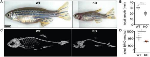
The mmp14a/b KO zebrafish recapitulate key aspects of the WS phenotype. (A) Gross anatomy photographs of 3-month-old WT and mmp14a/b KO fish of respective average size; lateral view, anterior to the left. The phenotype of mmp14a/b KO fish includes a relatively small, up-tilted head with relatively large, protruding eyes and a short operculum. Limited field of view necessitated stitching of multiple photographs together, causing the vertical line in the images shown in (A). Scale bar equals 2 mm. (B) At 90 dpf, mmp14a/b KO fish have a significantly shorter total body length compared with WT fish (P < 0.0001). A minimum of 21 individuals per genotype was measured. (C) 3D reconstruction of µCT scans of 3-month-old WT and mmp14a/b KO fish; lateral view, anterior to the left. Compared with WT fish, the mmp14a/b KO fish have Weberian-prehemal hyperkyphosis. (D) The mmp14a/b KO fish have a reduced skull bone mineral density (BMD, P < 0.05), giving the appearance of missing skeletal elements in the shown 3D reconstruction (C). BMD was assessed for five individuals per genotype. The individuals imaged in (C) are different from the ones shown in (A). |
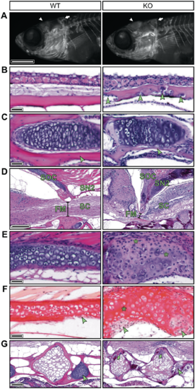
The mmp14a/b KO zebrafish have abnormal enchondral and membranous ossifying skull bones and Weberian vertebrae. (A) Fluorescence microscopy images of 30 dpf WT and mmp14a/b KO juveniles, whole mount stained for calcified bone with alizarin red; lateral view, anterior to the left. At 30 dpf, the frontal bones (arrowhead) and the supraoccipital bone (SOC, arrow) of mmp14a/b KO fish are shaped differently compared with age and size-matched (10.2 mm standard length) WT fish. (B–E) H&E stained mid-sagittal sections of 90 dpf fish; anterior to the left except for the mmp14a/b KO section shown in (E), which is rotated (anterior at the bottom) for clearer comparison with the corresponding WT section. At 90 dpf, the frontal bones (B) of mmp14a/b KO fish are irregularly thickened and contain cell clusters (arrowheads). The dentary bone (C) and SOC (E) of mmp14a/b KO fish contain a relative large amount of disorganized cartilage and small amounts of bone matrix (arrowheads). The SOC additionally shows cell-free areas that have lost basophilia, indicating lack of proteoglycans (E, asterisks). The SOC and second supraneural (SN2) form a sharp angle and are ventrally extended in mmp14a/b KO fish, impinging the spinal cord (SC) at the foramen magnum (FM, compare diameter indicated by black line in D). (F) Sagittal picrosirius red stained sections of the same fish as shown in (E) (same orientation as in (E) reveal the SOC of mmp14a/b KO fish lacks a collagen-rich peripheral bone matrix, but instead contains large cell-rich areas (arrowheads) as compared with WT fish. In contrast, cell-free regions in the cartilage core of mmp14a/b KO fish are relatively intensely stained. (G) H&E stained mid-sagittal sections (anterior to the left) demonstrating Weberian vertebral bodies of 90 dpf mmp14a/b KO fish are irregularly shaped and contain cell clusters (arrows), while the intervertebral cartilage is absent (arrowhead) compared with WT fish. Scale bar in (A) equals 1 mm, scale bars in (B), (C), (E) and (F) equal 20 µm, scale bar in (D) equals 200 µm, scale bar in (G) equals 100 µm. PHENOTYPE:
|

ZFIN is incorporating published figure images and captions as part of an ongoing project. Figures from some publications have not yet been curated, or are not available for display because of copyright restrictions. |
|
ZFIN is incorporating published figure images and captions as part of an ongoing project. Figures from some publications have not yet been curated, or are not available for display because of copyright restrictions. PHENOTYPE:
|
|
Unillustrated author statements PHENOTYPE:
|