
- Title
-
Genetic Requirement of talin1 for Proliferation of Cranial Neural Crest Cells during Palate Development.
- Authors
- Ishii, K., Mukherjee, K., Okada, T., Liao, E.C.
- Source
- Full text @ Plast Reconstr Surg Glob Open

Diagram depicting Talin1 (Tln1) protein with respect to the ECM (depicted by collagen), and the cell-surface (above). Tln1 links the actin cytoskeleton inside the cells to the ECM via the cytoplasmic domain of the β-subunit of integrins. Image of whole-mount embryo at 72 hpf shows the gene expression of tln1 in the craniofacial region during embryogenesis (below). Specific expression is observed in the palate (depicted by black arrowhead) and Meckel’s cartilage (depicted by black arrowhead and asterix). Scale bar: 500 μm.
EXPRESSION / LABELING:
|

Pulse-chase assay in Tg(sox10:kaede) transgenic to investigate palatal CNCC proliferation (above). Anterior (A) is up in all images. The distal tip of the palate is photoconverted from green to red at 60 hpf (pulse), in WT (left and above) and the mutant (left and below), with the contralateral side as control. Confocal images are taken after chase, at 72 hpf in both WT and tln1 (center). The palate is imaged in the green and the red channels in all instances. Green cells added onto the distal part of the palate during the chase depict the proliferative front, marked with white dotted line (center). Reduced cell proliferation is observed in tln1 mutants, (center and below) when compared with WT (center and above). EdU staining (green) reveals a proliferative front in both the WT and tln1 palate (red), marked by a white dotted rectangle (right). The proliferative cells are in yellow (overlap of green EdU+ and mCherry sox10+ CNCCs), depicted by white arrowheads. Scale bar: 100 μm. A, anterior; P, posterior.
|
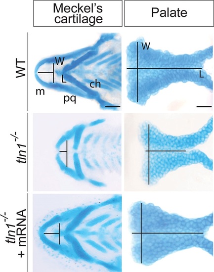
Alcian blue images of flat mounted Meckel’s cartilage and the palate of the WT (above), tln1 (center), and rescued mutants (below) at 4 dpf are shown. The length (L) of the Meckel’s cartilage is measured from the midline of the Meckel’s cartilage to the midline of an imaginary line drawn joining the joints between the Meckel’s cartilage and the palatoquadrate. Likewise the width (W) is across the joints between the Meckel’s cartilage and the palatoquadrate. The L of the palate is measured from the anterior to the posterior of the palate through its mid point, whereas W is measured as the maximum distance between the 2 lateral edges at the anterior most region. Tln1 mutants have a shorter Meckel’s cartilage (center and left) and palate (center and right). Injection of full-length tln1 mRNA partially rescues the phenotypes (below). Scale bars: 500 μm (Meckel’s cartilage) and 100 μm (palate). ch, ceratohyal; L, length; m, Meckel’s cartilage; pq, palatoquadrate; W, width.
PHENOTYPE:
|

Defects in craniofacial skeletal muscles in tln1 mutants. Confocal images of craniofacial skeletal muscles in Tg(mylz2:mCherry) transgenic reveal increased intermuscular tension in the craniofacial muscles in the mutants (left and center) when compared with WT (left and above). TEM images of the transverse section of the ih (depicted by white dotted line, above and center) show complete disruption of the sarcomere structure in the mutants, depicted by black arrowhead (right and center) when compared with WT (right and above). The Z-line and the A-and I-bands, observed in the WT (right and above) are completely absent in the mutants (right and center). Injection of full-length tln1 mRNA partially rescues the phenotypes as observed by decreased intermuscular tension in the craniofacial muscles in the rescued mutants (left and below). TEM images of the transverse section of the ih in the rescued mutants show reorganized sarcomere structure, similar to the WT (right and below). Scale bar: 500μm (Tg(mylz2: mCherry)) and 500nm (TEM). a, A-band; am, adductor mandibulae; i, I-band; ima, intermandibularis anterior; imp, intermandibularis posterior; s, sarcomere unit; sh, sternohyoideus.
|

ZFIN is incorporating published figure images and captions as part of an ongoing project. Figures from some publications have not yet been curated, or are not available for display because of copyright restrictions. EXPRESSION / LABELING:
|
|
ZFIN is incorporating published figure images and captions as part of an ongoing project. Figures from some publications have not yet been curated, or are not available for display because of copyright restrictions. PHENOTYPE:
|