
- Title
-
Expression of RPRM/rprm in the Olfactory System of Embryonic Zebrafish (Danio rerio).
- Authors
- Stanic, K., Quiroz, A., Lemus, C.G., Wichmann, I.A., Corvalán, A.H., Owen, G.I., Opazo, J.C., Concha, M.L., Amigo, J.D.
- Source
- Full text @ Front. Neuroanat.
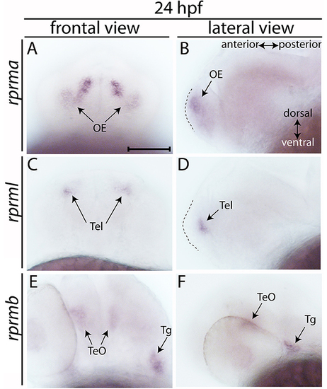
Expression of RPRM mRNA at 24 h post-fertilization (hpf). (A–E) The expression patterns of rprma/b and rprml were visualized by whole-mount in situ hybridization (WISH) during zebrafish early neuronal development. (A,C,E) Frontal views of the embryos heads, with dorsal to the top and ventral to the bottom (double arrow in B). (B,D,F) Lateral views of the embryos head with anterior to the left and posterior to the right (double arrow in B). (B,D) dashed areas represent the most anterior part of the embryo's head. At 24 hpf (A,B) rprma, (C,D) rprml, and (E,F) rprmb transcripts are located in neuronal cell populations such as: (A,B) olfactory placode (OP, black arrows), (C,D) telencephalon (Tel, black arrows), and (E,F) tectum opticum (TeO) and trigeminal ganglia (Tg), respectively. Scale bar in (A): 100 μm. EXPRESSION / LABELING:
|

Expression of RPRM mRNA at 48 and 72 hpf in embryonic zebrafish. Expression of RPRM transcripts was examined using WISH in wild-type embryos. At 48 and 72 hpf, (A–D) rprma, (E–H) rprml and (I–L) rprmb transcripts are located in specific cell populations. (A,C,E,G,I,K) Dorsal views of the embryos head, with anterior to the top and posterior to the bottom. (B,D,F,H,J,L) Lateral views of the embryos head with dorsal to the top, ventral to the bottom (double arrow in D), anterior to the left and posterior to the right (double arrow in D). rprma is expressed in the (A–D) olfactory system (OS, black arrows), habenular commissure (hbc, white head arrow) and at 72 hpf in presumptive distal cranial sensory ganglia (CSG, doted circle in D). rprml is expressed in the (E–H) Tel (black and white arrows), (G) diencephalon (Dien) and (H) latero-ventral midbrain (vMb). rprmb is expressed in the (I) OS (black arrow), (J) Hb, trigeminal ganglion (Tg, J,L) and (K,L) TeO (black arrows). Scale bar in (A): 100 μm. EXPRESSION / LABELING:
|
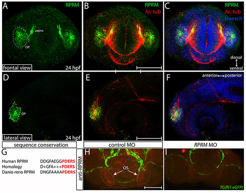
Expression of RPRM protein in the zebrafish olfactory system. RPRM protein localization was examined by immunofluorescence (IF) in wild-type embryos. At 24 h post-fertilization (hpf). (A–C) Frontal and (D–F) lateral views with dorsal to the top and ventral to the bottom (double arrows in C). (A,D) RPRM is expressed in the OP. The OP give rise to primary sensory neurons, and support basal cells of the OE. (B,E) Overlapped expression of RPRM with acetylated tubulin (Ac-tub) in the axons projecting to the presumptive olfactory bulb (OB) in the central nervous system (CNS). (C,F) Nuclei of the head cells are labeled by Hoechst staining. (G) Sequence conservation between human immunogen and zebrafish RPRM proteins. (H,I) RPRM protein expression is effectively blocked by antisense oligonucleotide MOs. (H,I) Frontal views of the head region in control-MO and RPRM MO-injected embryos by double immunofluorescence confocal microscopy at 48 hpf (with antibodies against RPRM and GFP). RPRM antibody labeled the olfactory system (OS, in red) in (H) MO-control injected embryos, but not in (I) RPRM MO-injected embryos. Scale bars in (B,E,H): 100 μm. |

Expression of RPRM in zebrafish olfactory epithelium. RPRM protein expression was analyzed by IF in wild-type embryos. (A–F) Frontal views with dorsal to the top and ventral to the bottom (double arrows in A,D). At 48 hpf (A–C) RPRM is expressed in the OE showing co-localization with Ac-tub. (B–C). At 72 hpf, (D–F) RPRM is expressed at the OE and co-localizes with olfactory sensory neurons (OSNs) which express Ac-tub (F–F′) and some of the axons projecting to the OB, also is present in the supraorbital neuromasts (SO) located bilaterally (dotted circles in D, F). (D′–F′) inset magnification at the OE showing RPRM and Ac-tub expression in OSNs (yellow square and arrows). Scale Bars in (A,D,D′): 100 μm. EXPRESSION / LABELING:
|

Expression of RRPM in zebrafish tectum opticum. Whole-mount immunofluorescence showing RPRM staining in a representative zebrafish embryo at 3dpf. (A–C) Dorsal view with anterior to the top, posterior to the bottom (double arrows in C). Expression of RPRM is shown in green and Ac-tub is shown in red. Positive labeling is observed in the OE, supraorbital neuromasts (SO, brackets), cranial sensory ganglia (CSG, brackets) and TeO (dotted white areas in A–C). (D–F) Magnification of TeO area with anterior to the upper-left and posterior to the bottom-right (double arrows in D), showing optic neuropil area labeled bilaterally (dotted white areas in D) as well as projections extending radially toward the midline (red dotted area in E). (F) Merged image for (D,E). (A–F) All the panels represent z-stack projections. Scale Bars in (A,D): 100 μm. EXPRESSION / LABELING:
|

RPRM expression is conserved between zebrafish and mice. (A–I) Zebrafish RPRM protein expression, visualized by IF at 72 hpf, presents similar expression patterns when compared with murine RPRM in comparable developmental stages. (A–C) Lateral view of a representative 72 hpf embryo by IF, showing RPRM (green) and Ac-tub (red) expression in the habenular commissure (hbc), OE, TeO, CSG, and hindbrain (Hb). Dorsal is show to the top and ventral to the bottom (double arrow in B) (D) Sagittal atlas image of E15.5 mouse embryo showing reference areas of the OS (OE, OB). (E) Nissl staining of sagittal section from E15.5 mouse embryo. (G,H) RPRM expression as detected by in situ hybridization (ISH) and (F) immunohistochemistry (IHC) for anti-GFP in the transgenic mice line TG(BAC-180MB-RPRM-EGFP). (F–I) RPRM expression pattern in TG(BAC-180MB-RPRM-EGFP) sections, showing low levels of RPRM gene products in the OB/OS (F) and high levels of expression in the OE (I). Scale Bars in (A): 100 μm, in (D): 1 mm. EXPRESSION / LABELING:
|