
- Title
-
A zebrafish model for ocular tuberculosis
- Authors
- Takaki, K., Ramakrishnan, L., Basu, S.
- Source
- Full text @ PLoS One
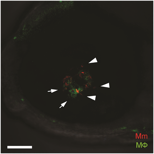
Confocal image of green-fluorescent macrophage aggregate surrounding red-fluorescent Mycobacterium marinum, located in the retinal parenchyma. The mycobacteria are intracellular within macrophages (arrows) as well as extracellular (arrowheads). Scale bar, 50 μm. PHENOTYPE:
|
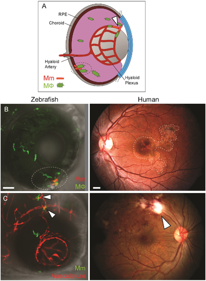
Anatomical localization of intraocular granuloma after M. marinum infection. (A) Schematic representation showing localization of granulomas near the retinal vasculature (arrowhead), and in retinal pigment epithelium-choroid complex (dotted circle). (B) Localization of intraocular granulomas (within dotted regions); in the outer eye of zebrafish larvae corresponding to the retinal pigment epithelium-choroid complex, and in the choroid in human ocular TB. (C) Localization of perivascular infection (arrowheads); as seen as bacterial aggregates in close association of blood vessels in zebrafish, and retinal periphlebitis associated with focal chorioretinitis overlying the blood vessel in human ocular TB. (B-C) Confocal images of ocular infection in Tg(mpeg:YFP) and Tg(kdrl:dsRed2) zebrafish, and fundus photographs of human ocular TB. Scale bars, 25 μm and 750 μm, respectively. PHENOTYPE:
|
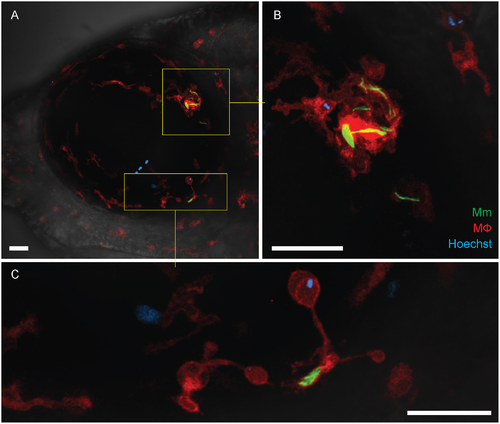
Recruitment of peripheral blood monocytes in granuloma formation. (A-C) Granuloma formation in Tg(mfap4:tdTomato) fish with red-fluorescent macrophages and infected with green-fluorescent M. marinum. Hoechst-positive (blue-fluorescent) peripheral blood monocytes which have been recruited into the infected eye are seen within the granuloma (inset and panel B) and in contact with a single infected macrophage (inset and panel C). The retinal microglia are incorporated into the granuloma and also dispersed uniformly within the ocular tissues. Scale bars, 30 μm. PHENOTYPE:
|