
- Title
-
The ADAMTS5 Metzincin Regulates Zebrafish Somite Differentiation
- Authors
- Dancevic, C.M., Gibert, Y., Berger, J., Smith, A.D., Liongue, C., Stupka, N., Ward, A.C., McCulloch, D.R.
- Source
- Full text @ Int. J. Mol. Sci.

Expression and silencing of adamts5 in zebrafish embryos. (A) ADAMTS5 expression in 8, 18 and 24 hpf wild-type embryos. Note strong early expression in the dorsal mesoendoderm (8 hpf, arrows) and variable expression ventrally (8 hpf, arrowhead), with later expression in the floor plate of the neural tube (18 and 24 hpf, arrows) and bilaterally in the prosencephalon (24 hpf, arrowheads). Asterisks = prosencephalon in no primary antibody control. Scale bar = 250 μm; (B) Schematic representation of the adamts5 gene structure targeted with antisense morpholino oligonucleotides (MO), and its subsequent splicing, indicating the primers used for RT-PCR and the size of the resultant products; (C) Reduced ADAMTS5 expression is seen in adamts5 AUG-MO injected embryos (asterisk) versus control (arrow) by whole-mount antibody labelling (left-hand panel) and Western blot (right-hand panel) showing the 120 kDa ADAMTS5 species (asterisk) with a region of the Coomassie blue stained gel shown below, demonstrating even loading; (D) RT-PCR of adamts5 mRNA obtained from 24 hpf embryos following injection of the adamts5 2/3-MO at the 1-cell stage, showing amplicons a and b (asterisk). β-actin was used as a house-keeping gene.
|

Notochord morphogenesis and muscle fiber formation is perturbed in adamts5 morphant embryos. (A) Expression of shh in the notochord of 18 hpf control (a,a′, arrows) and adamts5 morphant (b,b′, open arrowheads) embryos, with medio-lateral deviation in the adamts5 morphants (b,b′), with anterior indicated (*); (B) Expression of ntl in the notochord of 18 hpf control (a,a′, arrows) and adamts5 morphant (b,b′, open arrowheads) embryos, with medio-lateral deviation in the adamts5 morphants with anterior indicated (*); (C) Expression of myod in adaxial and paraxial mesoderm of 18 hpf control embryos (a,a′) and its perturbation in adamts5 morphants (b,b′, open arrowheads) with anterior indicated (*); (D) Quantitation of affected notochords in control and adamts5 morphant embryos demarcated by shh in Figure 2A; (E) Quantitation of affected notochords in control and adamts5 morphant embryos demarcated by ntl in Figure 2B; (F) Quantitation of embryos with perturbed myod expression in control and adamts5 morphant embryos demarcated in Figure 2C; (G) Double-transgenic Tg(acta1:lifeact-GFP)/Tg(acta1:mCherryCaaX) embryos, in which thin filaments are marked green and sarcolemma red, reveal loss of muscle integrity in 3 dpf adamts5 morphants. Muscle fibers of control injected larvae feature the typical striation of the myofibril and regular myofibers within chevron-shape somites, indicated by a dashed line (a). The boxed area in a is magnified in a′–a′′′. Myofibril striation is partially lost within adamts5 morphants (arrowhead in b′) and the sarcolemma of the myofibers disrupted (arrow in b′′). The boxed area in b is magnified in b′–b′′′. Scale bar = 50 μm.
|

Loss of paraxial mesodermal myod expression in adamts5 morphant embryos. (A) Expression of adaxial and paraxial myod in 12 hpf embryos injected with control MO (a, arrowheads), with adamts5 morphant embryos showing substantial loss of paraxial expression (b, open arrowheads), as well as mild loss of paraxial myod expression (b, open arrowheads). Rescue of paraxial myod expression in adamts5 morphants co-injected with mRNA encoding wild-type (c, arrows) or catalytically-inactive E411A (d, arrows) ADAMTS5. Control embryos injected with ADAMTS5 mRNA encoding wild-type ADAMTS5 show unaffected myod expression in paraxial mesoderm (e, arrows). Scale bar = 100 μm; (B) Quantitation of embryos showing present or absent myod patterning represented in (A).
|
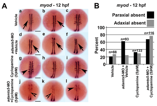
Combinatorial inhibition of Shh signaling and adamts5 disrupt paraxial and adaxial myod expression. (A) Adaxial and paraxial myod expression in 12 hpf embryos treated with vehicle control (a–c, arrows denote paraxial myod expression), vehicle + adamts5-MO (d–f, arrows denote absent paraxial myod expression), 5 μM cyclopamine (g–i, arrowheads represent similar paraxial myod staining compared to control group) and 5 μM cyclopamine + adamts5-MO (j–l, open arrowheads represent absent adaxial myod expression and arrowhead represents absent paraxial myod staining compared to adamts5 MO group). Scale bar = 200 μm; (B) Quantitation of embryos showing present or absent myod patterning represented in (A).
|

Combinatorial activation of Shh signaling and inhibition of adamts5 rescues paraxial myod expression. (A) Adaxial and paraxial myod expression in 12 hpf embryos treated with vehicle control (a–c, arrows), 10 μM Smoothened agonist (Smo agon.) (d–f, arrow/open arrowhead represent present/absent myod expression in paraxial mesoderm), vehicle + adamts5-MO (g–i, arrow/open arrowhead represents present/absent myod expression in paraxial mesoderm) and 10 μM Smo agon. + adamts5-MO (j–l, arrows indicate myod expression present in paraxial mesoderm). Scale bar = 100 μm; (B) Quantitation of embryos showing present or absent myod patterning represented in (A).
|

Expression of myod is altered in ADAMTS5 morphant embryos. (A) Expression of myod in paraxial mesoderm of 12 hpf control (arrows) and adamts5 exon 2/3 MO injected embryos. Note perturbation of expression in the adamts5 morphants (arrowheads). (B) Percent of embryos with perturbed myod expression in control and adamts5 morphant embryos represented in Supp Fig 1A. |