
- Title
-
Urban stormwater runoff negatively impacts lateral line development in larval zebrafish and salmon embryos
- Authors
- Young, A., Kochenkov, V., McIntyre, J.K., Stark, J.D., Coffin, A.B.
- Source
- Full text @ Sci. Rep.
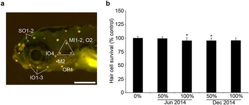
(a) Morphology of the head neuromasts in a larval zebrafish, visualized with DASPEI. Labels indicate the neuromasts that were quantified to assess hair cell damage. Scale bar = 250 µm. (b) Acute stormwater exposure does not kill lateral line hair cells. 5–6 dpf fish were exposed to stormwater for 24 hr (from June or December 2014 storm events), then hair cells were labeled with DASPEI. There is a slight but significant effect of stormwater (one-way ANOVA, F4,62 = 4.02, p = 0.006). Bonferroni-corrected post-hoc testing shows decreased hair cell survival for fish in the 100% June or 50% December groups (*p < 0.0001 for each). N = 13–14 fish per group, bars are + 1 s.d. |

Acute stormwater exposure decreases uptake of the transduction-dependent dye FM 1–43FX by hair cells. (a) Representative confocal images of neuromast IO3 from fish treated with no stormwater (0%), 50%, or 100% stormwater for 24 hr, then labeled with FM 1–43FX. The bar in the left panel = 10 µm and applies to all panels. (b) There is a significant reduction in FM 1–43FX fluorescence in stormwater-treated fish (one-way ANOVA, F2,25 = 19.81, p < 0.0001). Bonferroni-corrected post-hoc testing shows decreased fluorescence at 50% or 100% stormwater (****p < 0.0001 for each). Fluorescence was quantified in arbitrary units (A.U.). This experiment used stormwater from a June 2014 storm event. N = 7–11 fish per group, bars are + 1 s.d. |

Stormwater alters gross morphology of 4 dpf larvae exposed to runoff throughout development. (a) Untreated larvae (0% stormwater) showing normal development. (b-c) Examples of stormwater-treated larvae. (b) Some treated fish have not yet inflated their swim bladders (blue outlines). (c) Other treated fish show pericardial edema (blue arrows). Scale bar in a = 0.5 mm and applies to all panels. |

The Wnt activator Lithium chloride (LiCl) reduces the detrimental effects of stormwater treatment on the developing lateral line. All experiments were performed on Brn3c:mGFP fish treated from 4 hpf to 4 dpf. (a) Representative confocal images of the OP1 neuromast from zebrafish larvae raised in different stormwater concentrations, with and without the presence of LiCl. The scale bar in the upper left = 10 µm and applies to all panels. (b) Quantitative data showing significantly fewer hair cells in fish raised in stormwater from January 2016 (black bars), and prevention of hair cell loss when fish were raised in the same stormwater with LiCl present (gray bars). Similar to previous experiments, stormwater caused a significant decrease in hair cell number (two-way ANOVA, F2,72 = 10.15, p = 0.0001). LiCl-treated fish had significantly more hair cells than fish reared without LiCl (two-way ANOVA, F1,72 = 35.09, p < 0.0001). The interaction between the stormwater and LiCl treatments was also significant (two-way ANOVA, F2,72 = 3.819, p = 0.026). Asterisks indicate significant pairwise differences between -LiCl and + LiCl groups for a given stormwater concentration (****p < 0.0001). N = 10–16 fish per group treatment, bars are + 1 s.d. |

Developmental stormwater exposure does not alter cell proliferation within zebrafish neuromasts. Experiments were performed in Brn3c:mGFP fish, allowing for quantification of BrdU + /GFP + cells (hair cells). Fish were exposed to stormwater from 4 hpf – 4 dpf, and BrdU was pulsed at 32.5–34 hpf. (a) There was no change in the number of BrdU + hair cells in stormwater-treated embryos (black bars, two-way ANOVA, F2,44 = 1.701, p = 0.19). There was a significant increase in BrdU-labeled hair cells in LiCl-treated zebrafish (gray bars, two-way ANOVA, F1,44 = 13.72, p = 0.0006). Asterisks indicate significant pairwise differences between -LiCl and + LiCl groups for a given stormwater concentration. N = 7–13 zebrafish per group with exception of 50% stormwater + LiCl, where N = 4. Bars are + 1 s.d. (b) Representative confocal images showing BrdU labeling in the P2 neuromast of the trunk, with two hair cells double-labeled with GFP and BrdU (white arrows). Scale bar = 10 µm and applies to all panels. |
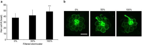
Filtered stormwater rescues neuromasts from the toxic effects of stormwater. All experiments were performed on Brn3c:mGFP fish treated from 4 hpf to 4 dpf. Filtered stormwater was diluted with EM; 0% therefore represents fish treated with clean water. (a) Fish exposed to filtered stormwater from January 2016 showed a significant increase in hair cell number in head neuromasts (one-way ANOVA, F2,63 = 8.092, p = 0.0007); post-hoc testing (Bonferroni-corrected) shows increase in the 100% filtered stormwater group (***p < 0.001). (b) Representative confocal images of the OP1 neuromast from zebrafish larvae raised in different filtered stormwater concentrations. N = 19–24 fish per group treatment, bars are + 1 s.d., scale bar = 10 µm and applies to all panels in (b). |
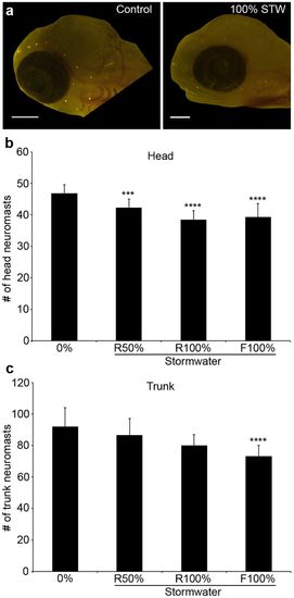
Stormwater exposure during development reduces the number of lateral line neuromasts in coho salmon. (a) Fluorescent images of DASPEI-labeled coho salmon embryos from a control fish (left) and a fish exposed to 100% stormwater (STW; right). Scale bars = 0.5 mm. (b) There are fewer head neuromasts in stormwater-exposed embryos (one-way ANOVA, F3,46 = 14.84, p < 0.0001). R = regular (unfiltered) stormwater, F = filtered stormwater. (c) There are also fewer trunk neuromasts in these embryos (one-way ANOVA, F3,46 = 8.33, p = 0.0002). For both (b) and (c), Bonferroni-corrected post-hoc analyses demonstrate significant differences from control embryos (***p < 0.001, ****p < 0.0001). N = 6–20 fish per treatment, bars are + 1 s.d. |