
- Title
-
Novel Animal Model of Crumbs-Dependent Progressive Retinal Degeneration That Targets Specific Cone Subtypes
- Authors
- Fu, J., Nagashima, M., Guo, C., Raymond, P.A., Wei, X.
- Source
- Full text @ Invest. Ophthalmol. Vis. Sci.
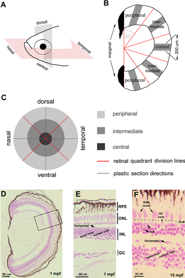
Linear cell densities of six categories of retinal cells were evaluated by JB4-Feulgen histology. (A) Zebrafish eyes were sectioned in either the nasal-temporal (anterior-posterior) or ventral-dorsal axis. (B) Each retinal section was partitioned by a vertical line to exclude the developing marginal region (black) from the differentiated retina, which was further divided into five regions each with angular subtense of 36 degrees. We counted nuclei in 200-μm linear segments (gray bars). (C) Spatial relationships among the nine sampled retinal regions. (D–F) JB4-Feulgen histology illustrates the localization and morphologies of the six retinal cell categories at 1 mpf (D, E; the inset box in D outlines the boundary of E) and 15 mpf (F). The dashed line indicates the location of the OLM. GC, ganglion cell layer; INL, inner nuclear layer; ONL, outer nuclear layer.
|

Topographical variations in planar cone densities and sizes. (A) Flat-mounted adult zebrafish retina viewed at a low magnification to illustrate the nine sampled regions. (B, C) Average planar cone densities in nine local regions sampled from retinas at 6 mpf, n = 3. (D–H) Representative images of ZO-1 immunolabeled apical cell profiles (at the OLM) from the nine local retinal regions. Arrows indicate UV cones; R, red cones; G, green cones; and B, blue cones. (G') The inset shows a higher magnification of the boxed region. Round profiles of rods are marked by arrowheads. (I) Abbreviations of retinal regions.
|

Secreted Crb2b-sfEX affects photoreceptor patterning and maintenance. (A) The expression patterns of Crb genes are highlighted with different colors superimposed on a transmission electron microscopic image of a tangential section through the photoreceptor layer. The Crb2b-sfEX transgene in pt108b is mostly expressed in blue cones; endogenous crb2a is expressed in all photoreceptors and Müller cells; endogenous crb2b is expressed selectively in RGB cones. (B) In flat-mount retinal preparations at 4 mpf, ZO-1 immunolabeled apical profiles of photoreceptors in pt108b retina form a mosaic pattern, but the regular organization is degraded by 18 mpf. (C, C') JB4-Feulgen staining of retinal cross sections from wild-type (WT) (C) and pt108 (C') illustrates severe reduction of RGB cones (arrows) in pt108b retina at 27 mpf, although rods and UV cone nuclei (arrowheads) are maintained. Dashed lines, OLM. (D, D') In WT retina (D) at 27 mpf Zpr1, immunolabeled green and red cones have elongated nuclei (arrows), but in pt108 (D'), the nuclei of the few remaining Zpr1-labeled cones are less elongated (arrow). UV opsin immunoreactivity (asterisks) verified the UV cone identity of nuclei (arrowheads) that are abnormally positioned apical to the OLM. DAPI (4′,6-diamidino-2-phenylindole), nuclear stain. Dashed lines, OLM. (E, E') ZO1 immunostaining (arrows) revealed the OLM, which is positioned apical to the UV cone nuclei in WT (E, arrowheads) but basal to UV cone nuclei in pt108b (E', arrowheads). UV cone outer segments were visualized by UV opsin immunostaining (asterisks).
|
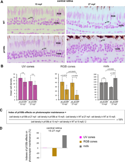
Secreted Crb2b-sfEX promotes RGB cone degeneration and rod overpopulation. (A) JB4-Feulgen histology illustrates nuclear morphologies and distributions of photoreceptors in the central area of WT and pt108b retinas at 15 mpf and 27 mpf. (B) Histograms of average linear densities of photoreceptors in central retina of WT and pt108b at 15 and 27 mpf. Mean ± 1 SD, n = 5. (C) Formula for the photoreceptor maintenance index. (D) In the central retina in pt108b, the photoreceptor maintenance indexes show loss of RGB cones, excess rods, and no effect on UV cones.
|

Planar cone density analysis confirmed secreted Crb2b-sfEX produces age-related cone loss. (A–E'') Comparison of ZO-1 immunolabeled apical cone profiles in representative regions from 26-mpf WT, 18-mpf pt108b, and 20-mpf pt108b retinas.
PHENOTYPE:
|

Secreted Crb2b-sfEX induces apoptosis in cones. Retinal flat-mount preparations double-immunolabeled with anti-Crb2a (green) and the apoptotic marker, anti-active caspase 3 (magenta). (A–A''', B–B''') Cells labeled with anti-active caspase 3 were in the photoreceptor layer, as indicated by anti-Crb2a label. (A–A'') and (B–B'') represent 2D planar views, and (A''') and (B''') include 3D-reconstructed “cut views” (in the xz and xy planes) at the level of the OLM, indicated by the white cross hairs, demonstrating that apoptotic and photoreceptor-specific labels are in the photoreceptor layer. (C–C''') Active caspase 3–labeled photoreceptors were not observed in WT retinas.
|